What is an impacted canine and how common is it?
An impacted canine is a permanent canine that has not come through at its normal time and stays in the bone, usually in the upper jaw. According to the guideline of the Royal College of Surgeons of England (RCS), the upper canine is, after the lower wisdom tooth, the tooth most often impacted, and the problem affects about 1.5% of people; a Cochrane review gives 1 to 3%.
A canine becomes impacted much more often on the palate side than on the cheek side, and canines lying towards the cheek more often come through on their own, without treatment. The cause is not fully understood and is probably genetic: a canine impacted in the palate is more common in families and in children with missing or small lateral incisors, no crowding and delayed development of the teeth (RCS, 2022).
What are the first signs that a canine is not coming through properly?
The first sign is the absence of the typical bulge above the baby canine. Most permanent canines that erupt normally can be felt with a finger on the cheek side at the age of 10 to 11, and a canine that erupts later than about 12.3 years in girls and 13.1 years in boys is considered late (RCS, 2022).
To detect an impacted canine in a child early, the RCS guideline recommends that from the age of 8 the dentist looks at and feels the canine area every year. X-rays before the age of 10 or 11 usually add little. When to take a child to an orthodontist for the first time is described in a separate article; the American Association of Orthodontists (AAO) recommends a first check-up by the age of 7 at the latest.
- The canine cannot be felt on the cheek side at the age of 10 to 11.
- The canines on the two sides come through unevenly, or the bulge can be felt on one side only.
- The lateral incisor is tilted or unusually mobile.
- The lateral incisor is missing or clearly small, which is linked to a higher risk.
What are the risks of a canine that stays in the bone?
The main risk is resorption, that is, the dissolving of the roots of the neighbouring incisors. In one study using CT, resorption was found in 66.7% of lateral incisors next to an impacted canine, although it was not always clinically significant (RCS, 2022).
Resorption of the incisor roots rarely begins after the age of 14 and most often appears between 11 and 12, which is why early diagnosis matters. In adults the crown of the canine itself may be resorbed, reported in up to 14% of cases, while cystic change is rare.
How the dentist locates the canine: X-rays and CBCT
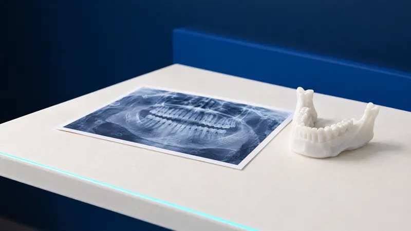
The position of an impacted canine is usually established on two X-rays taken with the X-ray tube in different positions, using the parallax effect: the apparent shift of the canine’s image relative to the incisors shows which side of the arch it lies on. According to the RCS, a horizontal shift of the tube gives a more reliable result than a vertical one.
Small field of view cone beam computed tomography (CBCT) allows the canine to be located precisely and the incisor roots to be assessed, but it involves a higher radiation dose than ordinary X-rays. That is why British and European guidelines reserve it for selected cases where ordinary X-rays are not enough to plan treatment, and the British Orthodontic Society stresses that it is not taken routinely. The doses of each examination are compared in the article on CBCT and panoramic X-rays.
What are the treatment options for an impacted canine?
The choice depends on age, the position of the canine, the condition of the neighbouring teeth and whether the patient wants to wear fixed braces and is able to. The RCS strongly recommends seeking a specialist’s opinion before starting on any of these paths.
| Option | When it is considered | What it involves |
|---|---|---|
| Monitoring | No resorption or other changes, good contact between the neighbouring teeth | Regular clinical examination, no active treatment |
| Removing the baby canine | Age about 10 to 13, canine not too severely displaced | Extraction of the baby canine and an X-ray assessment after 12 months |
| Exposure and fixed braces | Canine can be brought into place, patient ready to wear braces | Exposure surgery, then fixed braces, usually for 2 to 3 years |
| Removing the permanent canine | No consent to treatment, early incisor resorption, canine too far out of position | Surgical removal of the canine, sometimes moving a premolar into its place |
| Canine transplantation | When other treatment has failed or is not an option | Moving the patient’s own canine to the correct place in the arch |
Removing the baby canine: when does it help?
Removing the baby canine is an interceptive treatment that improves the position of the permanent canine in some children. The RCS provides for it in carefully selected children aged about 10 to 13 whose canine is not severely displaced, with better results when there is no crowding; if no improvement is visible on an X-ray after 12 months, other treatment is considered.
The first study of this method reported 78% of canines returning to a normal eruption path, and a later one 62%. A 2021 Cochrane review covering 4 studies and 199 children, however, rated the evidence as very uncertain: extraction may increase the proportion of canines that come through on their own, but it cannot be ruled out that there is no difference (RR 2.87; 95% CI 0.90 to 9.23).
The closer the canine lies to the middle of the palate, the less often it comes through on its own, with or without treatment. Removing two baby teeth instead of one did not give better results, and parents should know that exposure and treatment with braces may be needed despite the extraction.
Exposure of an impacted canine and treatment with braces: what does it involve?
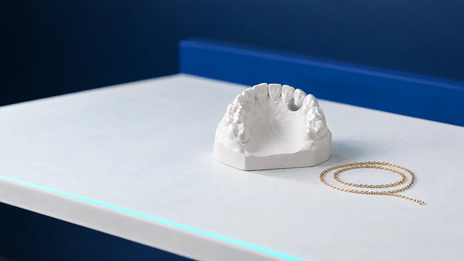
Exposure of an impacted canine is a surgical procedure after which the orthodontist gradually brings the canine into place with fixed braces. A Cochrane review describes two techniques: closed, in which an attachment with a small chain is bonded to the canine and the mucosa is stitched back over it, and open, in which the tissue over the crown is removed so that the canine stays visible.
Both techniques may be equally effective (3 studies, 146 participants, low-quality evidence), and individual studies found no differences in pain, appearance, gum condition or treatment time. Treatment of an impacted canine with braces itself usually takes 2 to 3 years of wearing fixed braces (Parkin et al., 2017).
The RCS lists the conditions: the patient is ready to wear fixed braces, is well motivated and has healthy teeth, and the canine does not lie so far out of position that bringing it into place is impossible, for example right next to the midline, above the root tips of the neighbouring teeth or horizontally.
Impacted canine in an adult: what changes?
In an adult, an impacted canine can also be brought into place with braces, but the chances are lower and treatment takes longer. In a study of 19 adults with a mean age of 28.8 years, the success rate was 69.5%, while in a group of teenagers with similarly difficult canines treatment succeeded in every case; all the failures involved people over 30, and the adults needed more time and more visits (Becker and Chaushu, 2003).
The RCS explains this by the risk, rising with age, of the canine fusing with the bone and of resorption of its crown, and considers adolescence the optimal time for exposure. When an adult does not want or is unable to have the canine brought into place, its removal is considered; if the gap will then need filling, for example with an implant, it is worth planning treatment together with a prosthodontist from the start.
How it works at DentalBlue
At DentalBlue in Białystok, the X-rays needed to locate the canine, including panoramic X-rays, cephalometric X-rays and CBCT scans, are taken in the clinic’s own radiology unit. Orthodontic treatment, with fixed braces or Clear Aligner trays, is planned digitally on the basis of cephalometric analysis, and surgical procedures are performed under local anaesthesia or, for qualified patients, under general anaesthesia. In the clinic’s price list, “Konsultacja ortodontyczna” (orthodontic consultation) costs 300 zł, “Odsłonięcie zęba zatrzymanego” (exposure of an impacted tooth) 500 zł and “Operacyjne usunięcie zęba całkowicie zatrzymanego” (surgical removal of a fully impacted tooth) 1 000 zł. The dentist sets the final scope and cost after the examination, and prices may change.
Frequently asked questions
Can an impacted canine come through on its own?
Sometimes, yes. Canines lying towards the cheek more often come through without treatment, and some palatal canines change their path after the baby canine is removed. The closer the canine lies to the middle of the palate, the lower the chance of this, which is why the decision is based on X-rays and successive check-ups.
At what age is it best to treat an impacted canine?
Removing the baby canine is considered at around 10 to 13, and according to the RCS exposure followed by treatment with braces is best carried out during adolescence. In adults treatment is possible, but studies show lower effectiveness and a longer treatment time.
Does an impacted canine need to be removed if it causes no problems?
Not always. The RCS accepts leaving the canine in place when there is no resorption of the neighbouring roots or other changes, and the neighbouring teeth are in good contact in the arch. Such a canine does, however, need regular check-ups at the dentist; the guidelines do not specify how often to take X-rays if there are no symptoms.
How much does exposure of an impacted canine cost?
In the clinic’s price list, “Odsłonięcie zęba zatrzymanego” (exposure of an impacted tooth) costs 500 zł, and “Miniimplant ortodontyczny” (orthodontic mini-implant), if the treatment plan includes one, 700 zł. On top of this comes treatment with fixed braces, priced after an orthodontic consultation (300 zł). The dentist sets the final scope and cost, and prices may change.
Does exposure of a canine hurt?
At DentalBlue the procedure is performed under local anaesthesia or, for qualified patients, under general anaesthesia. Studies comparing exposure techniques found no differences in the pain experienced; isolated complications were reported, such as infection or pain when the chain pushes through the gum of the palate.
Sources
- Royal College of Surgeons of England: Management of the Palatally Ectopic Maxillary Canine (2022)
- Benson PE et al., Cochrane: interceptive treatment of palatally displaced canines in children (2021)
- Parkin N et al., Cochrane: open versus closed exposure of palatal canines (2017)
- Becker A, Chaushu S, Am J Orthod Dentofacial Orthop: treatment of impacted canines in adults (2003)
- AAO: Why Kids Should See an Orthodontist by Age 7 (2026)
An informational article. It does not replace a consultation, and treatment decisions are made during a visit.