How does a panoramic X-ray differ from a CBCT scan?
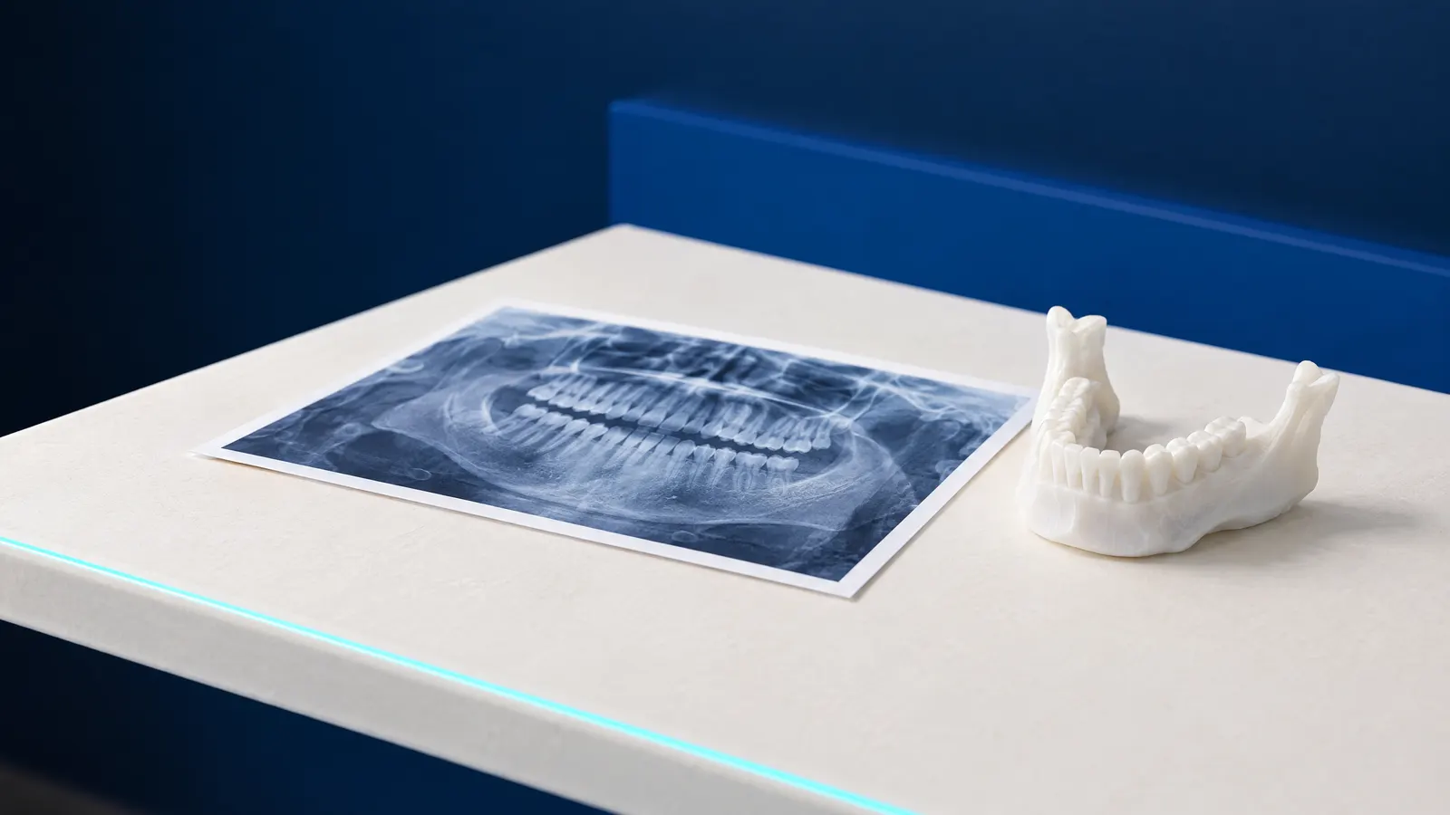
A panoramic X-ray and a CBCT scan differ above all in the number of dimensions. A panoramic X-ray (orthopantomogram) is a single flat image showing all the teeth, the roots and the bones of the upper and lower jaw, laid out like a map. Cone beam computed tomography (CBCT) takes a series of images around the head and assembles them into a three-dimensional image, which the dentist views slice by slice, in any plane.
A panoramic X-ray answers the question of what is happening in the whole mouth. CBCT answers the question of exactly what a specific site looks like: how much bone there is for an implant, where the nerve canal runs in the lower jaw, how many canals a root has and whether it is fractured. On a flat image structures overlap; on a CT scan they can be separated.
CBCT is not the same as the computed tomography done in hospital. A dental cone beam scanner covers a smaller area and usually gives a clearly lower dose than medical CT of the jaws.
| Feature | Panoramic X-ray | CBCT scan |
|---|---|---|
| Image | Flat, all the teeth and both jaws at once | Three-dimensional, a chosen area or both jaws |
| Typical dose | 2.7 to 24.3 µSv | Small and medium field: 11 to 674 µSv, median 61 µSv |
| What it is used for | Overview of the mouth, treatment plan, assessment of wisdom teeth | Implants, difficult canals, impacted teeth close to the nerve |
| When it is taken | Often as the first image for a new problem | For a specific indication, not as a screening test |
| Price in the clinic's price list | 120 zł | 300 zł one jaw, 350 zł both |
How much radiation does a dental CT scan give compared with a panoramic X-ray?
A dental CT scan usually gives a dose several times higher than a panoramic X-ray, but both are measured in microsieverts (µSv), thousandths of a millisievert. According to European guidelines, the typical effective dose of a panoramic X-ray is 2.7 to 24.3 µSv, of small and medium field CBCT 11 to 674 µSv (median 61 µSv), and of large field CBCT 30 to 1,073 µSv (median 87 µSv) (European Commission, Radiation Protection 172, 2012).
The spread is wide, because the dose depends on the machine, the field size and the settings. A 2015 meta-analysis reports for adults an average of 84 µSv with a small field, 177 µSv with a medium field and 212 µSv with a large field, and for children an average of 103 µSv with a small field (Ludlow et al., 2015). That is why the smallest practical field is chosen, covering only the area needed.
For comparison: natural background radiation in Poland gives an average of 2.55 mSv a year, about 7 µSv a day (Poland's National Atomic Energy Agency, 2025). We calculated the last column of the table from this value; it is an approximation for typical doses, not a measurement of a specific machine.
| Examination | Typical dose | Equivalent in natural background radiation in Poland |
|---|---|---|
| Intraoral X-ray | below 1.5 µSv | less than 1 day |
| Cephalometric X-ray | below 6 µSv | under 1 day |
| Panoramic X-ray | 2.7 to 24.3 µSv | up to about 3.5 days |
| Small or medium field CBCT | 11 to 674 µSv, median 61 µSv | median: about 9 days |
| Large field CBCT | 30 to 1,073 µSv, median 87 µSv | median: about 12 days |
| Medical CT of the jaws | 280 to 1,410 µSv | about 40 to 200 days |
When is a panoramic X-ray enough?
A panoramic X-ray is enough when the dentist needs an overview of the whole mouth: at the first visit, when planning treatment of many teeth, before prosthetic treatment, and when assessing wisdom teeth and the buds of permanent teeth in children. One image shows larger cavities, lesions at the root tips, impacted teeth and bone loss.
For wisdom tooth removal, a panoramic X-ray is the first-choice examination. According to European recommendations, CBCT is not a routine examination here: it is taken only when the surgeon has a specific question that a flat image cannot answer, for example when the panoramic X-ray shows the root in close contact with the nerve canal in the lower jaw (EADMFR, 2019).
For details of a single tooth, such as assessing the canals during treatment or decay between teeth, the dentist usually turns to an intraoral X-ray. It covers a small area but shows it more sharply, at a very low dose.
When is a CBCT scan needed?
A CBCT scan is needed when the answer depends on the third dimension and a flat image cannot provide it. It is not a screening test: the American endodontic and oral radiology societies state that CBCT should not be used routinely, only for a specific indication (AAE and AAOMR, 2015).
A CT scan shows more, but more findings do not always mean less risk. In a study of 379 lower wisdom teeth assessed on both types of image, changes around the tooth were seen more often on CBCT, and on that basis more teeth would have been put forward for removal (Matzen et al., 2017), so the decision on the scan rests with the dentist, who knows what question it is meant to answer.
- Implant planning: bone height and width, the position of the nerve canal in the lower jaw and of the floor of the maxillary sinus.
- Root canal treatment with unclear symptoms, complex root anatomy or calcified canals.
- Failed root canal treatment and the planning of retreatment or an apicoectomy.
- Suspected vertical root fracture, dental injuries and root resorption.
- An impacted tooth whose root touches the nerve canal on the panoramic X-ray, if a 3D image would change the surgical plan.
Is a dental CT scan safe?
A dental CT scan taken for a justified indication gives a dose usually equivalent to days or weeks of natural background radiation. Safety in radiology, however, is not only about the dose being small but about every image being justified: it is taken when the result can change a treatment decision, and with as low a dose as possible.
That is why there is no single answer to how often a panoramic X-ray or a CT scan can be taken. The indication is decided by the dentist on the basis of the examination, earlier images and the treatment plan. If you have recent images from elsewhere, bring them to the visit, because they sometimes make a repeat unnecessary.
Are panoramic X-rays and CBCT covered by the NFZ?
A panoramic X-ray is covered by the NFZ, with restrictions, while CBCT is not included in the NFZ list of dental services. Adults are covered for a panoramic X-ray for the purposes of root canal treatment, and every patient before prosthetic treatment, no more than once every 5 years (Ministry of Health regulation, Dz.U. 2025 item 615).
The current text of the regulation requires a panoramic X-ray for an adult to be referred by a specialist or a doctor in specialist training, and the referring doctor justifies the need for the examination in the referral. Children and young people aged 5 to 18 are covered for a panoramic X-ray when it is medically justified, once every 3 years.
Intraoral X-rays under the NFZ are limited to 2 per calendar year for adults and 5 for children and young people. The law is stated as at 12 October 2026.
How much do a dental CT scan and a panoramic X-ray cost?
The price of a dental CT scan depends mainly on its extent: whether the image covers one jaw or both. A panoramic X-ray has one fixed price, and intraoral X-rays are charged per image.
In the clinic's price list, a CT scan of the upper or lower jaw costs 300 zł and of both jaws 350 zł. A panoramic X-ray is 120 zł, a sectional panoramic X-ray 60 zł, an intraoral X-ray 60 zł, a cephalometric X-ray 120 zł and a sinus X-ray 100 zł. A panoramic X-ray is also part of the first visit, with or without pain. The dentist decides which examinations are needed, and prices may change.
What happens during the scan and how do you prepare?
A dental X-ray, including a CBCT scan, needs no special preparation and you do not have to fast, because it is taken without contrast. The examination is short, and the patient stands or sits still in the machine.
- Remove earrings, glasses, hair clips, chains and removable dentures, because metal casts shadows on the image.
- Tell the team if you are pregnant or cannot rule it out: the dentist will judge whether the image is needed now or can wait.
- Bring earlier images if you have them, including on a disc or as a file.
- Keep still and breathe calmly during the exposure, because movement blurs the image and may mean a repeat.
How it works at DentalBlue
DentalBlue in Białystok has its own radiology unit, where CBCT scans, panoramic X-rays, cephalometric X-rays, sinus X-rays and intraoral X-rays are taken, so there is no need to go anywhere else for an image. A panoramic X-ray is part of the first visit, with or without pain. Implants are planned on a CBCT image from this unit, and CT is also used in root canal treatment under a microscope and before surgical procedures, when the dentist considers it necessary.
Frequently asked questions
Is CBCT the same as a CT scan?
CBCT is a type of computed tomography in which the X-ray beam is cone-shaped and covers a chosen area. Typical CBCT doses in dentistry are lower than those of medical CT of the jaws, for which European guidelines give 280 to 1,410 µSv.
Does a panoramic X-ray show tooth decay?
Larger cavities, yes, but a panoramic X-ray is not precise enough to detect early decay between teeth. That is the job of bitewing intraoral X-rays, which show a small area more sharply; one study gives a combined 5.0 µSv for four such images (Ludlow et al., 2008).
Do you need a referral for a dental CT scan?
The indication for a CT scan is decided by the dentist, who knows what question it is meant to answer. Under the NFZ, CBCT is not included in the list of dental services, and a panoramic X-ray for an adult needs a referral from a specialist or a doctor in specialist training.
Can a child have a dental CT scan?
Yes, when there is an indication, for example an injury or an impacted tooth. In children it is particularly important to limit the field to the area needed; a meta-analysis gives an average of 103 µSv for children with a small field (Ludlow et al., 2015). Children from the age of 5 are covered by the NFZ for a panoramic X-ray once every 3 years when it is justified.
Does a panoramic X-ray have to be taken every year?
There is no rule that a panoramic X-ray is taken every year. An image is taken when its result could change treatment, and the interval depends on the state of the teeth and the plan. The NFZ funds a panoramic X-ray for adults once every 5 years in specific situations, but that is a funding limit, not a medical recommendation.
Sources
- European Commission: Radiation Protection 172, Cone Beam CT for Dental and Maxillofacial Radiology, SEDENTEXCT guidelines (2012)
- Ludlow JB et al., Dentomaxillofac Radiol: CBCT doses, meta-analysis (2015)
- Ludlow JB et al., JADA: doses of intraoral and panoramic X-rays (2008)
- Państwowa Agencja Atomistyki: Raport roczny (2025)
- AAE and AAOMR: joint position statement on CBCT in endodontics (2015)
- Matzen LH, Berkhout E, Dentomaxillofac Radiol: EADMFR position statement on CBCT for third molars (2019)
- Matzen LH et al., Dentomaxillofac Radiol: third molars on panoramic X-ray and CBCT (2017)
- Dz.U. 2025 poz. 615: rozporządzenie MZ w sprawie świadczeń gwarantowanych z zakresu leczenia stomatologicznego (2025)
An informational article. It does not replace a consultation, and treatment decisions are made during a visit.