What types of dental X-ray are there?
A dental X-ray is the everyday term for an intraoral X-ray, an image taken with a plate or sensor placed inside the mouth. There are several types, and the dentist chooses the one that answers a specific question: what is happening around a root, whether there is decay between teeth or where an impacted tooth lies.
According to the American medical encyclopaedia MedlinePlus, a periapical X-ray shows one or two whole teeth from crown to root, while a bitewing image shows the crowns of the upper and lower teeth together as the patient bites on a special holder. The table sets out the types of image and their typical uses.
| Type of X-ray | What it shows | Typical use |
|---|---|---|
| Periapical X-ray | One or two teeth from crown to root tip and the surrounding bone | Root canal treatment, toothache, abscess, injury, planning an extraction |
| Bitewing X-ray | The crowns of upper and lower back teeth together, with the teeth biting | Decay between teeth and beneath fillings, bone level |
| Occlusal X-ray | A larger part of the arch on a plate resting against the chewing surfaces | Position of impacted teeth, injuries and tooth development in children |
| Full-mouth series | All the teeth in a series of intraoral X-rays | Extensive oral disease or extensive previous treatment |
| RVG (radiovisiography) | Any of the above images recorded with a digital sensor instead of film | A prompt on-screen image, for example during root canal treatment |
What is RVG, and how does it differ from a film X-ray?
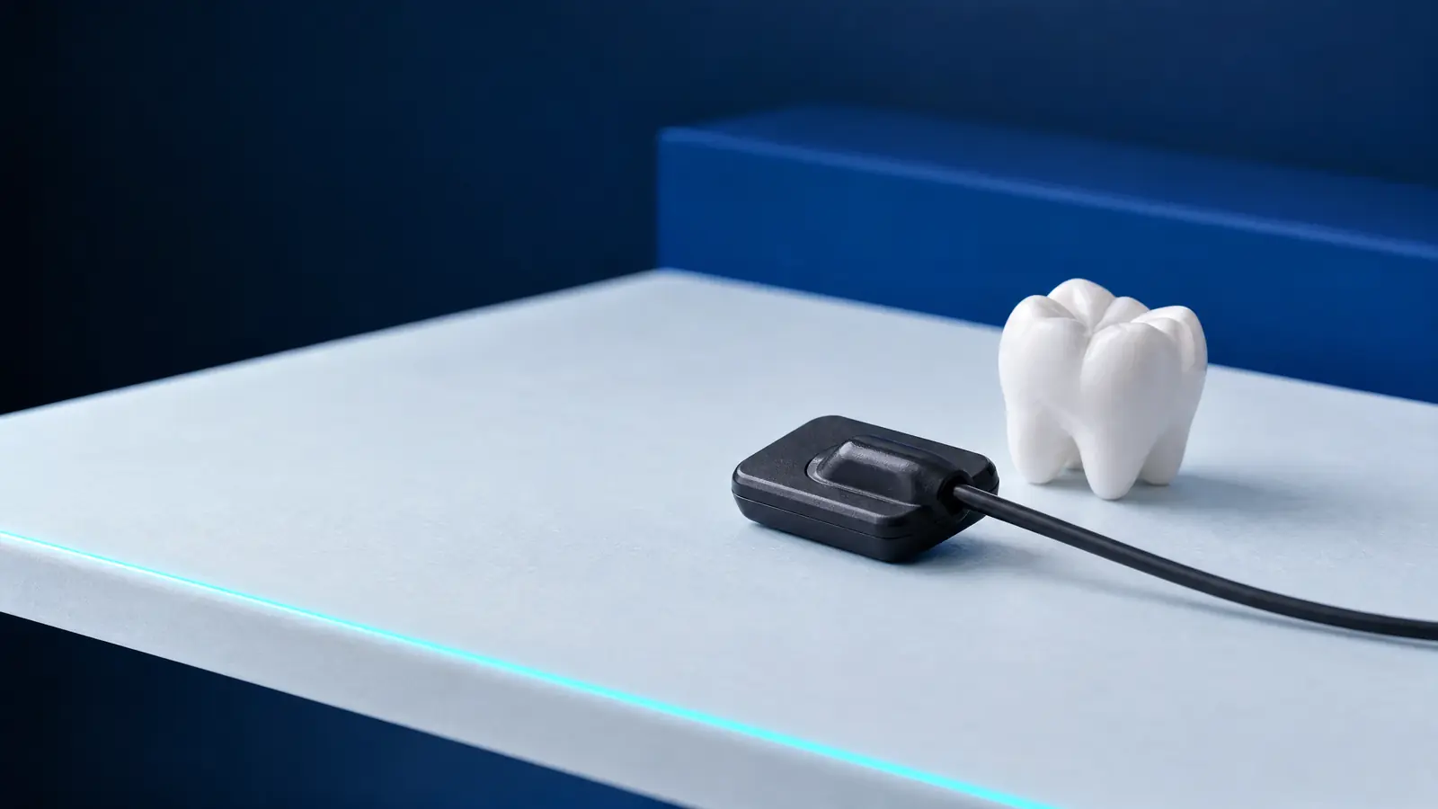
RVG, or radiovisiography, is a digital dental X-ray taken with an electronic sensor instead of film. The term even appears in legislation: the regulation on dental services funded by the NFZ, Poland’s National Health Fund, lists “aparat RTG do zdjęć zębowych lub radiowizjografia” (a dental X-ray machine or radiovisiography) as equipment (t.j. Dz.U. 2025 poz. 615).
The 2012 ADA and FDA guidelines state that digital imaging can reduce the dose by 40 to 60%. With direct sensors, the image appears on the monitor within a few seconds, while phosphor plates must first be scanned.
Beam shape also affects the dose: according to the same guidelines, a rectangular collimator reduces it by as much as fivefold compared with a circular one. The Polish regulation requires a voltage of 60 to 70 kV for intraoral X-rays and rectangular collimation with a sensor holder where possible (§ 5 ust. 3, t.j. Dz.U. 2025 poz. 1288).
When does a dentist take a dental X-ray?
A dentist takes a dental X-ray when the result could change a treatment decision, rather than routinely at every visit. The ADA and FDA guidelines state that images are ordered after an examination and medical history when the additional information will affect patient care. In a study of 490 patients cited in the guidelines, this approach reduced the number of X-rays by 43% without a clinically significant increase in missed disease.
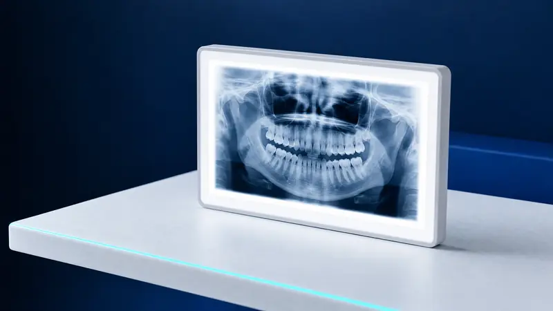
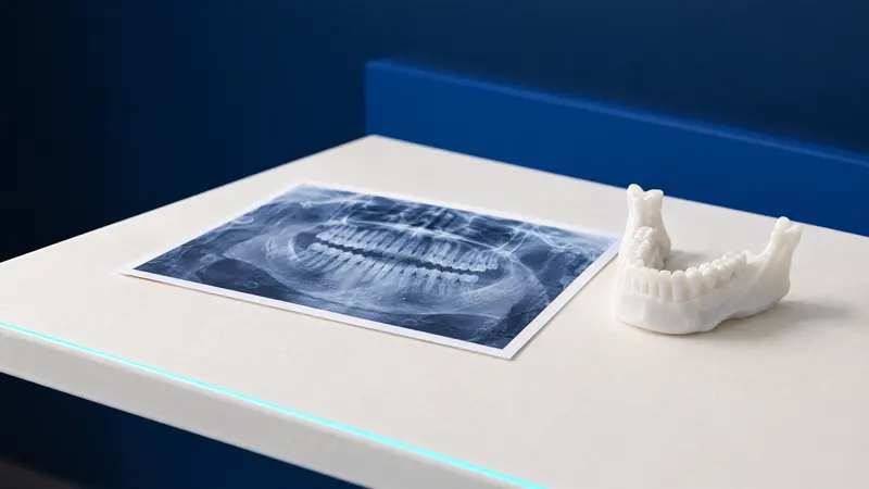
According to these guidelines, intraoral X-rays are more accurate than panoramic images for assessing dental injuries, root shape, root resorption and pulp disease. A panoramic X-ray, by contrast, provides an overview of the whole mouth, as explained in the article on panoramic dental X-rays. Typical situations in which a dentist uses a dental X-ray include:
- Root canal treatment: images before treatment, during treatment, after the canals have been filled and at follow-up.
- Toothache, swelling or a suspected abscess, when the root and the bone around its tip need to be assessed.
- Suspected decay between teeth that cannot be seen in a mirror or examined with a probe.
- Dental injury, a suspected root fracture or resorption, meaning loss of root tissue.
- Planning a tooth extraction or checking after a surgical procedure.
How often are bitewing X-rays taken?
The interval between bitewing X-rays depends on the risk of decay, rather than a fixed schedule. For patients attending check-ups, the 2012 ADA and FDA guidelines give the intervals listed below for situations in which the surfaces between teeth cannot be seen or examined with a probe.
These are recommendations, not limits: the guidelines stress that decay risk changes over time, so the interval between X-rays needs to be adjusted accordingly. The dentist sets it after examining you. How often to attend check-ups themselves is explained in the article on dental check-ups.
- Children and teenagers with decay or an increased risk: every 6 to 12 months.
- Adults with decay or an increased risk: every 6 to 18 months.
- Children without decay or an increased risk: every 12 to 24 months.
- Teenagers without decay or an increased risk: every 18 to 36 months.
- Adults without an increased risk: every 24 to 36 months.
- People with no teeth: X-rays are not recommended if there are no signs of disease.
Do you need a referral for a dental X-ray?
An intraoral dental X-ray does not require a referral if it is taken using a machine intended exclusively for such images. This is an exception to the general rule that examinations using ionising radiation require a referral or an order (§ 3 ust. 1 i 3 of the Minister of Health’s regulation, t.j. Dz.U. 2025 poz. 1288). A separate condition applies to the NFZ-funded service covering up to 5 intraoral images for children and young people under 18: the referring doctor must justify the need for the examination in the referral, including the number of images.
Not needing a referral does not remove the need for justification. The Atomic Law Act requires the expected benefit of an examination to outweigh the possible harm to health (art. 33c, t.j. Dz.U. 2026 poz. 1), so the dentist examining you decides whether an X-ray is needed. Panoramic X-rays and CT scans require a referral or an order. Legal status as of 24 October 2026.
How much radiation does a dental X-ray involve?
According to the European Radiation Protection 172 guidelines, a single intraoral image typically involves less than 1.5 µSv with rectangular collimation. A 2008 study by Ludlow and colleagues reports 5.0 µSv for four bitewing images and 34.9 µSv for a full-mouth series taken with a sensor or fast film and rectangular collimation.
In the same study, the full-mouth series using older, slower film and circular collimation gave 388 µSv, more than 11 times as much, illustrating how much depends on equipment and technique. The article on CBCT and panoramic X-rays compares the doses from dental X-rays, panoramic images and CBCT scans.
X-ray machines in dental surgeries are subject to oversight: permission to use X-ray machines for medical purposes is issued by the state provincial sanitary inspector (art. 5 ust. 4 of the Atomic Law Act). In Podlaskie province, this is the inspector based in Białystok (as of 24 October 2026).
Dental X-rays during pregnancy: are they allowed, and is an apron used?
Dental X-rays are taken during pregnancy when needed for diagnosis or treatment: the American Dental Association (ADA) considers dental X-rays safe at every stage of pregnancy (page updated on 14 July 2025). Poland’s Atomic Law Act gives pregnant women special protection, and art. 33e ust. 2 requires information on pregnancy or breastfeeding to be obtained where it is relevant to the procedure.
The regulations require particular attention above all to examinations involving the abdomen and pelvis, whereas a dental X-ray covers a small area of the head (art. 33e ust. 4). Treatment during this period is discussed further in the article on dental treatment during pregnancy.
In Poland, personal protective shields are used for dental X-rays, especially thyroid protection, provided this does not reduce the image’s diagnostic value (§ 5 ust. 1 pkt 3 of the regulation, t.j. Dz.U. 2025 poz. 1288). The ADA no longer recommends abdominal or thyroid shielding for dental X-rays, including during pregnancy, but Polish regulations apply in a Polish surgery.
How much does a dental X-ray cost, and what does the NFZ cover?
The cost of dental X-rays depends on the number of images and whether they form part of treatment. In the clinic’s price list, “Zdjęcie wewnątrzustne” (intraoral X-ray) costs 60 zł, “RVG w chirurgii” (RVG for surgery) 100 zł, and “Pakiet zdjęć RVG do leczenia endodontycznego: przed, w trakcie i po leczeniu oraz kontrolne” (RVG package for root canal treatment: before, during and after treatment, plus follow-up images) 200 zł. The dentist sets the final scope and cost at the consultation, and prices may change.
Under the NFZ, every patient is entitled to up to 2 intraoral X-rays per calendar year in conjunction with other services. Children and young people up to the age of 18 are entitled to up to 5 images per year in conjunction with an examination or root canal treatment (Minister of Health’s regulation, t.j. Dz.U. 2025 poz. 615). For the children’s service, the referring doctor must justify the need for the examination in the referral, including the number of images. Legal status as of 24 October 2026.
Dental X-rays at DentalBlue
At DentalBlue in Białystok, intraoral X-rays, panoramic X-rays and CBCT scans are performed in the clinic’s own radiology unit, and the team includes radiographers. For root canal treatment, the clinic’s price list provides a package of RVG images taken before, during and after treatment, and at follow-up. The dentist decides on the number and type of images after the examination.
Frequently asked questions
Does a dental X-ray hurt?
No. According to MedlinePlus, the X-ray itself causes no discomfort. For some people, the plate or sensor in the mouth triggers a gag reflex, which calm, deep breathing through the nose usually helps relieve.
What is a periapical dental X-ray?
A periapical X-ray shows one or two teeth from the crown to the root tip, together with the surrounding bone. It is taken, for example, before root canal treatment, for toothache or before a tooth extraction.
Can a child have a dental X-ray?
Yes, when the result is needed for treatment. The Atomic Law Act provides special protection for people under 16, and the ADA and FDA guidelines link the frequency of X-rays in children to decay risk. The NFZ funds up to 5 intraoral images per year for children in specified circumstances, on a referral that justifies the examination and states the number of images.
Can a dental X-ray show decay between teeth?
Yes, this is primarily what bitewing images are used for. The ADA and FDA guidelines identify them as the most effective method for detecting cavities on the surfaces where teeth touch. According to these guidelines, some such cavities can only be detected on an X-ray.
Why does the dentist take several X-rays during root canal treatment?
Images at successive stages show the tooth before treatment, during treatment and after the canals have been filled, while a follow-up image allows the dentist to assess healing of the bone around the root later on. The dentist chooses the number of images to suit the course of treatment, and a digital RVG sensor allows immediate viewing.
Sources
- MedlinePlus (U.S. National Library of Medicine): Dental x-rays (2026)
- ADA and FDA: Dental Radiographic Examinations, Recommendations for Patient Selection and Limiting Radiation Exposure (2012)
- European Commission: Radiation Protection 172, CBCT in dentistry, SEDENTEXCT guidelines (2012)
- Ludlow JB et al., JADA: doses from intraoral and panoramic X-rays (2008)
- ADA: Oral Health Topics, Pregnancy (2025)
- Dz.U. 2025 poz. 1288: rozporządzenie MZ w sprawie warunków bezpiecznego stosowania promieniowania jonizującego dla wszystkich rodzajów ekspozycji medycznej (2025)
- Dz.U. 2026 poz. 1: ustawa Prawo atomowe, consolidated text (2026)
- Dz.U. 2025 poz. 615: rozporządzenie MZ w sprawie świadczeń gwarantowanych z zakresu leczenia stomatologicznego (2025)
An informational article. It does not replace a consultation, and treatment decisions are made during a visit.