What is tooth decay, and how does it develop?
Tooth decay is a chronic disease of the hard tissues of the tooth in which the bacteria of dental plaque (biofilm) turn sugars from food and drink into acids, and the acids gradually dissolve the enamel and dentine. In its fact sheet of March 2025, the World Health Organization (WHO) lists as the main causes a constant, high intake of free sugars, too little exposure of the teeth to fluoride, and plaque that is not removed by brushing.
Decay is not a one-way process. After a sugary meal, acid washes minerals out of the enamel, and between meals saliva and fluoride partly restore them; a review in Nature Reviews Disease Primers (Pitts et al., 2017) describes decay as a dynamic, sugar-driven disease of the biofilm, in which the fate of the tooth depends on the balance between the loss and the regaining of minerals.
What matters is not only the amount of sugar but also how often it reaches the mouth. The WHO recommends that free sugars, meaning those added to food and those naturally present in honey, syrups and fruit juices, provide less than 10% of dietary energy, and preferably less than 5%, which is about 25 g a day.
How common is tooth decay in adults in Poland?
Tooth decay affects almost all adults in Poland. In the most recent nationwide survey, carried out under a Ministry of Health programme and summarised in 2021 by the Medical University of Warsaw, 98.7% of people aged 35 to 44 had signs of decay, meaning cavities, fillings or teeth extracted because of it (data from 2017).
The same survey also brings good news: in nearly 50% of people in this age group no active decay was found, and the result improved markedly compared with 2010. Among people aged 65 to 74 (data from 2019), 18.8% had no teeth at all, compared with 40.8% ten years earlier.
These are still the most recent national figures, because nationwide monitoring was not continued after 2020. Globally, the WHO, drawing on Global Burden of Disease 2021 data, regards untreated decay of the permanent teeth as the most common health condition in humans.
What does tooth decay look like at each stage?
Tooth decay develops in stages: first acid washes minerals out from beneath the surface of the enamel and a dull, chalky white spot forms, then the surface collapses and a cavity, that is a hole in the tooth, appears, and an untreated lesion reaches through the dentine to the pulp. Each stage calls for a different approach, as the table shows.
On the contact surfaces between the teeth, early lesions are usually not visible to the naked eye. The dentist assesses their depth on an X-ray, most often a bitewing, and that assessment determines whether treatment without drilling is enough.
| Stage | What you see or feel | What usually helps |
|---|---|---|
| White spot in the enamel | A dull, chalky spot, usually painless | Fluoride toothpaste, fluoride varnish, check-ups |
| Lesion between the teeth without a cavity | Nothing; the lesion shows up only on an X-ray | Fluoride, resin infiltration or sealing |
| Cavity in the enamel and dentine | A hole, dark discolouration, sensitivity to sweet things | A filling after the decay has been removed |
| Deep decay close to the pulp | Pain with cold or sweet things that passes quickly | A filling that preserves the pulp, sometimes in stages |
| Inflamed or dead pulp | Pain may be spontaneous; a dead pulp may cause no pain | Pulp assessment; depending on the diagnosis, pulp-preserving or root canal treatment |
| Tooth destroyed down to the root | Remnants of the crown, sometimes swelling of the gum | Assessment of whether the tooth can be restored, or extraction |
What are the symptoms of tooth decay, and what does a black spot on a tooth mean?
At first, tooth decay often causes no symptoms at all, which is why many cavities are found only at a check-up. The NHS, the UK’s public health service, lists as later signs toothache, sensitivity to hot, cold or sweet things, and white, brown or black spots on a tooth.
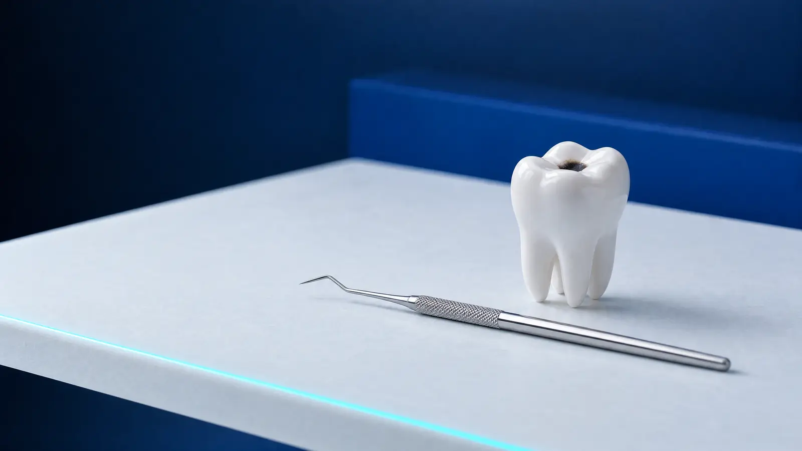
A black spot on a tooth, most often in a groove of a molar, does not always mean active decay. The Scottish SDCEP guidance on assessing the activity of carious lesions stresses that the colour of a lesion is not a reliable indicator of its activity, because some arrested lesions are dark and some are light; a hard, smooth and shiny surface suggests an arrested lesion, and a soft, matt one an active lesion.
Only a dentist can assess hardness and texture, and until the examination SDCEP advises treating every lesion as active. A dark spot may also be a stain from coffee, tea or tobacco; the list sets out the symptoms worth seeing a dentist about.
- Pain when eating sweet, cold or hot food that keeps coming back in one place.
- A hole or a rough spot where food gets caught.
- A white, brown or black spot that grows larger over time.
- A dark shadow under the enamel or at the edge of an old filling.
- Spontaneous or night-time pain, which may mean an inflamed pulp and needs a prompt visit.
Can tooth decay be stopped?
Tooth decay can be stopped: an early lesion in the enamel, and even an initial lesion in the dentine, can be arrested without drilling, as can selected lesions with a cavity, especially on roots, under a dentist’s supervision. Arresting the disease does not, however, restore lost tissue, and the dentist decides on the treatment. The NHS says that a dentist can reverse early decay with fluoride products, for example a varnish or a mouthwash.
Fluoride toothpaste is the foundation. The UK guidance Delivering better oral health (DBOH, updated on 10 September 2025) recommends that adults brush at least twice a day, including before bed, with a toothpaste containing 1,350 to 1,500 ppm fluoride, and spit out the foam without rinsing the mouth with water. For adults at increased risk, DBOH recommends fluoride varnish twice a year and, with active decay, considering a toothpaste with 2,800 or 5,000 ppm fluoride prescribed by the dentist.
For lesions between the teeth, micro-invasive treatment helps. A Cochrane review (Dorri et al., 2015) showed that resin infiltration or sealing clearly reduced the chance of non-cavitated lesions progressing compared with fluoride varnish alone or flossing instruction (odds ratio 0.24; 7 studies, 602 lesions); this applied to lesions reaching at most the outer third of the dentine.
Fluoride will not close a cavity that has already formed. The NHS says that a hole in a tooth usually needs a filling, and fluoride and good hygiene then protect the edges of the filling and the remaining teeth.
Secondary decay and root decay: why do they affect adults?
Secondary decay is decay that develops next to a filling or a fissure sealant; this is how a review prepared for the European cariology organisations ORCA and EFCD defines it (Askar et al., 2020). The authors note that gaps at the edge of the filling, the individual patient’s risk of decay and the experience of the dentist placing the filling matter more than the material itself.
Decay under a filling is, alongside fracture, one of the two main reasons why fillings fail. In an analysis of 12 studies with at least 5 years of follow-up (Opdam et al., 2014), fillings in back teeth in people at high risk of decay failed at a rate of 3.2% a year, compared with 1.2% in people at low risk. Askar et al. add that secondary decay, once detected, is treated by repairing the damaged part or by replacing the whole filling, and that poorly validated methods of detecting it can lead to unnecessary replacements.
Root decay affects exposed root surfaces, most often where the gums have receded. A meta-analysis by Pentapati et al. (2019) estimates its prevalence at 41.5%, or about 4 in 10 adults, and a review by Heasman et al. (2017) indicates that active root lesions can be arrested with fluoride varnishes or high-fluoride toothpaste, and that those the patient cannot clean well can be filled using the least invasive methods possible.
How is tooth decay treated at each stage?
Treatment of tooth decay depends on whether the tooth surface is intact or a cavity has formed, and on how close the lesion comes to the pulp. A lesion without a cavity is treated with fluoride and hygiene, and between the teeth also with resin infiltration; a cavity usually needs restorative treatment, that is a filling, the types of which are described in a separate article on fillings.
With deep decay, the aim is to keep the pulp alive. In its 2019 position statement, the European Society of Endodontology (ESE) recommends, for teeth with reversible pulpitis, selective removal of decay, in one or two stages, so as not to expose the pulp. According to the 2023 ESE guideline, with pulpitis and spontaneous pain the dentist may consider root canal treatment or, in selected cases, a pulpotomy, a treatment that keeps the living part of the pulp. With a dead pulp, root canal treatment is usually needed, and when the tooth cannot be saved, extraction.
On the NFZ, Poland’s National Health Fund, adults are entitled to treatment of superficial decay, which under the regulation also covers treatment of initial decay, and to fillings of cavities on 1, 2 and 3 surfaces. The funded materials are glass ionomer cements and, in the teeth from canine to canine, chemically cured composite. For most adults the NFZ funds root canal treatment of incisors and canines; women who are pregnant or in the postpartum period are also entitled to it in the back teeth, under the terms of the guaranteed benefits (legal status as of 24 October 2026).
How can adults prevent tooth decay?
Tooth decay is prevented by limiting how often you have sugary food and drinks, giving the teeth fluoride every day and removing plaque regularly. DBOH recommends that adults reduce the amount and frequency of sugary products, and the interval between check-ups is set by the dentist according to individual risk, as described in a separate article on dental check-ups.
The risk increases with a dry mouth, because saliva neutralises acids and restores minerals. Dry mouth can be a side effect of medicines: a review by Wolff et al. (2017) identified 56 substances with strong evidence of this effect. Do not stop taking medicines on your own; talk to the doctor in charge of your treatment instead.
- Brush your teeth twice a day, including before bed, with a fluoride toothpaste of 1,350 to 1,500 ppm.
- After brushing, spit out the toothpaste and do not rinse your mouth with water.
- Have sweets and sugary drinks with meals rather than in small amounts throughout the day.
- Clean between your teeth every day with floss or interdental brushes.
- Tell your dentist about a dry mouth, receding gums and any new medicines.
How it works at DentalBlue
At DentalBlue in Białystok, decay is diagnosed by the dentist on the basis of an examination and, when needed, X-rays from the clinic’s own radiology unit: an intraoral X-ray or a panoramic X-ray. A “Wizyta bez bólu” (visit without pain, 300 zł) includes a panoramic X-ray, a consultation, an examination and a written treatment plan. In the clinic’s price list, “icon-infiltracja żywicą” (Icon resin infiltration) costs 350 zł, “Fluoryzacja” (fluoride treatment) 100 zł and “Wypełnienie jednopowierzchniowe” (single-surface filling) 300 zł. The final scope and cost are set by the dentist at a consultation, and prices may change. The clinic is open Monday to Friday from 8:00 to 20:00 and on Saturdays from 8:00 to 16:00.
Frequently asked questions
Does tooth decay always hurt?
No. The NHS stresses that decay may cause no symptoms at all at first, and pain usually appears when the cavity is deep or the pulp has become inflamed. That is why a dentist finds many lesions at a check-up or on an X-ray before the tooth starts to hurt.
Can a hole in a tooth heal by itself?
No. Fluoride and good hygiene can stop a lesion from progressing, but the tooth cannot rebuild a cavity that has already formed. According to the NHS, a hole in a tooth usually needs a filling, and if left untreated it can reach the pulp.
Does decay between the teeth show up on an X-ray?
Yes, and often only there, because the contact surfaces cannot be seen when the teeth are examined by eye. The UK FGDP criteria recommend bitewing X-rays every 6 months at high risk of decay, every 12 months at moderate risk and about every 2 years for adults at low risk.
Can tooth decay be treated without drilling?
Often yes, particularly if the lesion has not yet formed a cavity: fluoride is used, and between the teeth resin infiltration or sealing, which according to a 2015 Cochrane review slowed the progression of lesions better than fluoride varnish alone. Under a dentist’s supervision, selected lesions with a cavity, mainly on roots, can also be arrested without drilling. Arresting decay does not rebuild lost tissue, and many cavities need a filling.
Do honey and fruit juice harm teeth as much as sugar?
The WHO classes the sugars in honey, syrups and fruit juices as free sugars, just like sugar added to food, so as far as tooth decay is concerned they count in much the same way. The WHO recommends that free sugars provide less than 10% of dietary energy, and preferably less than 5%.
Sources
- WHO: Oral health, fact sheet (2025)
- Ministerstwo Zdrowia, WUM: Choroba próchnicowa i stan tkanek przyzębia populacji polskiej (tooth decay and periodontal status of the Polish population), surveys from 2016 to 2019 (2021)
- NHS: Tooth decay (2025)
- OHID: Delivering better oral health, chapter 2, summary guidance tables (2021, updated 2025)
- Cochrane: Dorri M et al., micro-invasive treatment of decay on contact surfaces (2015)
- International Endodontic Journal: Duncan HF et al., ESE S3-level clinical practice guideline for the treatment of pulpal and apical disease (2023)
- SDCEP: Prevention and Management of Dental Caries in Children, assessing carious lesion activity
- ELI: Rozporządzenie MZ w sprawie świadczeń gwarantowanych z zakresu leczenia stomatologicznego, t.j. Dz.U. 2025 poz. 615
An informational article. It does not replace a consultation, and treatment decisions are made during a visit.