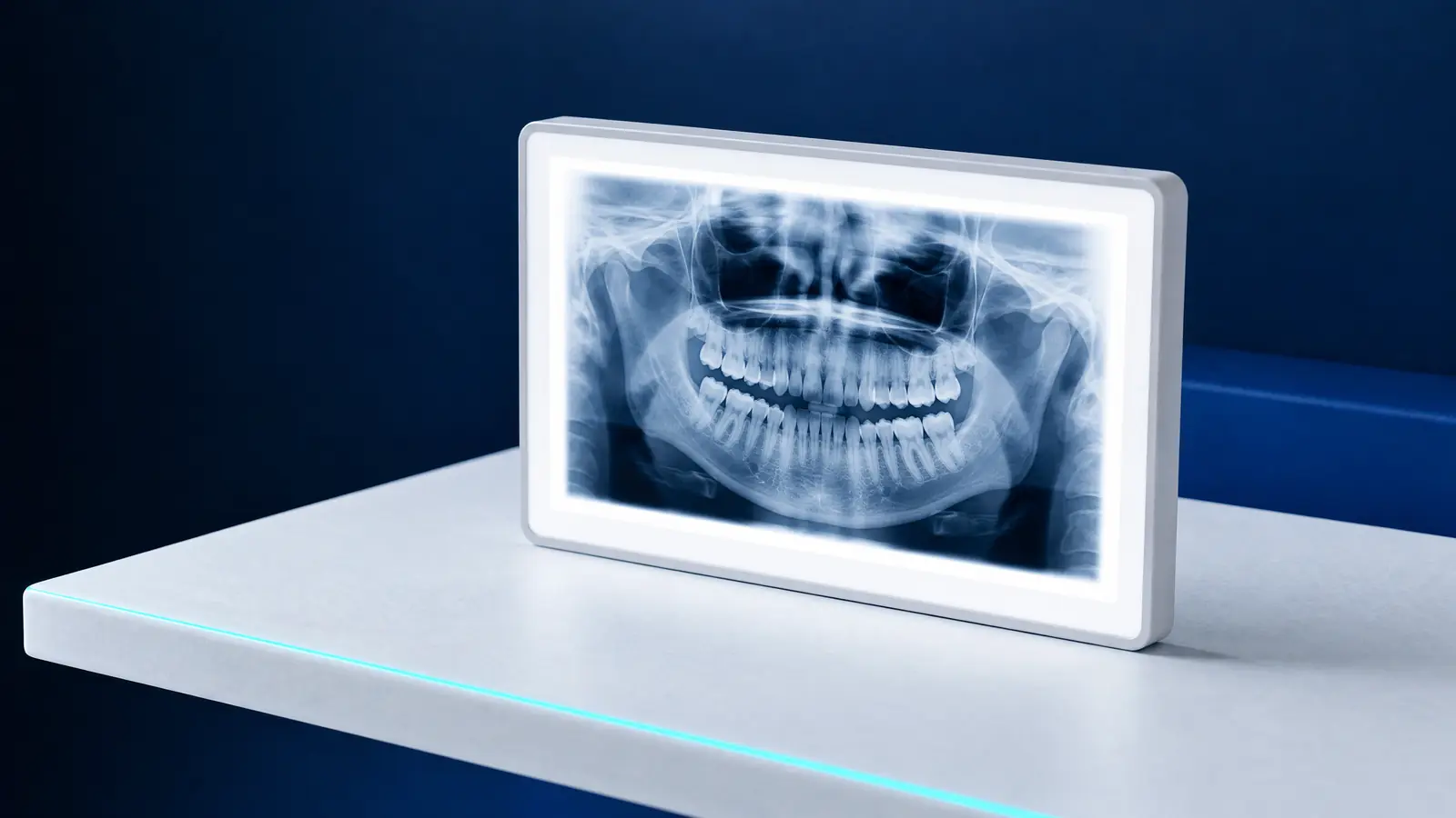
Dental X-rays in Białystok: which images are taken at a practice?
Dental X-rays cover several different examinations; the clinician chooses the one that will answer a specific question. An intraoral radiograph shows one or several teeth in detail, a panoramic radiograph shows the whole mouth in one image, cephalometry provides a side-view head image for orthodontic measurements, and CBCT provides a three-dimensional image of a selected area.
A review in ‘Insights into Imaging’ (Suomalainen et al., 2015) describes panoramic and intraoral radiographs as basic dental examinations, and CBCT as an examination for more demanding cases. The table gives typical doses from the European Commission’s 2012 figures, cited in that review. The dose for an individual examination depends on the equipment and settings.
| Examination | What it shows | Typical effective dose |
|---|---|---|
| Intraoral radiograph (RVG) | One or several teeth, their roots and surrounding bone | below 1.5 µSv |
| Panoramic radiograph | All teeth, roots and the upper and lower jawbones | 2.7 to 24.3 µSv |
| Cephalometric radiograph | Side-view head image for orthodontic measurements | below 6 µSv |
| Dentoalveolar CBCT (field up to 10 cm high) | 3D image of a selected part of the jaws | 11 to 674 µSv, median 61 µSv |
| Craniofacial CBCT (field over 10 cm) | 3D image of a larger area of the head | 30 to 1 073 µSv, median 87 µSv |
Do dental X-rays require a referral?
Panoramic radiographs, cephalometric radiographs and CBCT scans require a referral or order, even when you pay privately. Section 3 of the ‘Rozporządzenie Ministra Zdrowia w sprawie warunków bezpiecznego stosowania promieniowania jonizującego dla wszystkich rodzajów ekspozycji medycznej’ (Minister of Health regulation on safe use of ionising radiation for all types of medical exposure), in its 2025 consolidated text, requires radiation examinations to be based on a referral or order. Exemptions include dental intraoral radiographs taken with equipment dedicated solely to that purpose.
A doctor or dentist can refer you: ‘Prawo atomowe’ (Atomic Law), in its 2026 consolidated text, includes both in its definition of a referring clinician. The referral contains the purpose and justification, preliminary diagnosis and information needed to perform the examination. At a practice with its own equipment, the dentist examining you may order the image.
If another clinician has referred you, bring the referral, because the purpose and preliminary diagnosis determine the imaging scope. For example, the scan field is selected according to the area relevant to the clinician’s question, so a referral without an examination purpose makes planning difficult.
An intraoral image not requiring a referral does not mean imaging on demand. ‘Prawo atomowe’ requires every radiation examination to be justified by the expected benefits outweighing possible harm to health, and a referral to be based on the clinician’s conviction that the result will help diagnosis or treatment (Article 33c). Legal position as at November 2026.
How much do dental X-rays cost in Białystok?
DentalBlue’s published price list has a separate ‘X-rays’ category, and some images are also included in other items. The table brings together all imaging-related items; names follow the price list.
If this is your first visit for a new problem, check the first-visit items: they already include a panoramic X-ray. Prices may change, and the clinician decides the type and number of images after examining you.
| Price-list item | Price | What it includes |
|---|---|---|
| ‘Panoramic radiograph’ | 120 zł | Image of all teeth and jawbones |
| ‘Sectional panoramic radiograph’ | 60 zł | Image of a selected arch segment |
| ‘Intraoral radiograph’ | 60 zł | Image of one or several teeth |
| ‘Cephalometric radiograph’ | 120 zł | Side-view head image for orthodontic measurements |
| ‘Sinus radiograph’ | 100 zł | X-ray of the sinuses |
| ‘CBCT scan, upper or lower jaw’ | 300 zł | CBCT of one jaw |
| ‘CBCT scan, both jaws’ | 350 zł | CBCT of both jaws |
| ‘RVG X-ray in surgery’ | 100 zł | Digital tooth X-ray during a surgical procedure |
| ‘RVG X-ray package for root canal treatment: before, during, after and follow-up’ | 200 zł | Images at successive stages of root canal treatment |
| ‘First visit without pain’ | 300 zł | Panoramic X-ray, consultation, examination and written treatment plan |
| ‘First visit in pain’ | 340 zł | Panoramic X-ray, consultation, examination and pain relief |
Panoramic radiograph: what does it show and what happens?
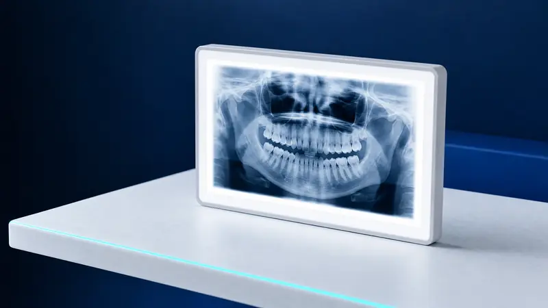
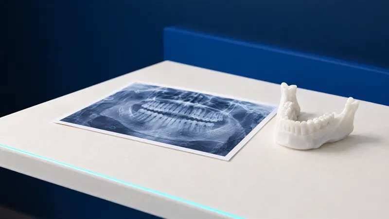
A panoramic radiograph is a single two-dimensional image of the whole mouth: all teeth and the upper and lower jawbones. RadiologyInfo, run by American radiological societies, describes it as an initial assessment examination used, among other things, to plan dentures, orthodontic appliances, extractions and implants. It does not provide fine detail of individual teeth or soft tissues.
According to RadiologyInfo, panoramic imaging helps identify conditions including advanced periodontal disease, jawbone cysts and tumours, impacted teeth including wisdom teeth, temporomandibular joint disorders and sinusitis. Where detail of one tooth is needed, the clinician adds an intraoral radiograph; where the answer depends on a third dimension, a scan.
No special preparation is needed. RadiologyInfo advises removing jewellery around the head, glasses and other metal objects, and always mentioning a possible pregnancy. The examination proceeds as follows:
- Your head is positioned against supports and you hold a bite block in your mouth to position the teeth correctly.
- The machine’s arm rotates around your head, usually for 12 to 20 seconds; you must stay still because movement blurs the image.
- The equipment can be adjusted for someone standing or sitting in a wheelchair.
- The examination does not hurt, and no sensor or film is placed in your mouth.
RVG tooth X-ray: when is a small image enough?
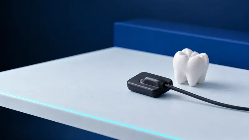
An intraoral radiograph is taken with a sensor or plate inside the mouth and shows a selected tooth in greater detail than a panoramic image. RVG, or radiovisiography, is this type of image recorded digitally using a sensor instead of film; the term even appears in the NFZ dental benefits regulation as practice equipment.
A clinician usually chooses a small image when the question concerns one site: a root during root canal treatment, a tooth before extraction or a post-procedure check. DentalBlue therefore lists separate RVG items for surgery and root canal treatment alongside ‘Intraoral radiograph’.
Polish regulations also specify how these images are taken: at 60 to 70 kV and, where possible, with rectangular collimation and a sensor holder (section 5(3) of the regulation). These rules aim to limit the beam to the necessary area and keep the dose from a single image very low.
For people with a strong gag reflex, RadiologyInfo notes that a panoramic radiograph may be recommended instead of intraoral images, because nothing except the bite block needs to go in the mouth. Mention this problem before the examination.
Dental CBCT in Białystok: when is it needed?
Cone-beam computed tomography (CBCT) produces a three-dimensional image in which the clinician can examine the jaws layer by layer. The review by Suomalainen et al. (2015) recalls recommendations from the European SEDENTEXCT project: CBCT should not be routine, and the clinician should have a history, clinical examination findings and previous images beforehand. A scan is justified when two-dimensional images cannot answer the diagnostic question and 3D imaging will add important new information.
Typical situations include assessing bone before an implant, or a lower wisdom tooth whose root appears to contact the nerve canal on a conventional image, once a decision to remove it has already been made. The same review recommends the smallest possible scan field, and Polish regulations require dental tomography to be limited to the clinically relevant area (section 5 of the regulation).
The same review also cites the recommendation against routine large-field CBCT in orthodontic diagnosis. It may be justified for complex skeletal problems, particularly those treated with both orthodontics and surgery. If a scan is proposed, ask what question it is intended to answer.
CBCT is not listed among NFZ dental benefits, so it is performed privately. The article on when to choose CBCT or a panoramic radiograph compares them, including their doses.
Cephalometric radiographs in Białystok: why does an orthodontist need one?
A cephalometric radiograph is a side-view head X-ray on which the orthodontist measures the relative positions of the jaws and teeth. It forms the basis of cephalometric analysis, the starting point for an orthodontic treatment plan. The typical dose in the European Commission’s figures is below 6 µSv.
DentalBlue lists ‘Cephalometric radiograph’ at 120 zł and, under orthodontics, ‘Treatment plan with cephalometric analysis’ at 380 zł. NFZ orthodontic benefits for children and young people include a cephalometric radiograph, with its analysis covered twice during the entire course of treatment where justified.
NFZ panoramic and dental X-rays in Białystok: what is covered?
NFZ dental X-rays are provided as part of treatment with a contracted dentist, rather than a standalone examination on request: the dental benefits regulation links them to other NFZ benefits. Adults are entitled to up to 2 intraoral radiographs per calendar year. Adults can have a panoramic radiograph for root canal treatment, and any patient before prosthetic treatment, once every 5 years, on referral from a dentist with a specialist qualification or undergoing specialist training.
Children and young people aged 5 to 18 can have a panoramic radiograph once every 3 years in medically justified cases, and up to 5 intraoral radiographs per year alongside an examination or root canal treatment; the referring clinician must justify the need, including the number of images, in the referral. Children undergoing orthodontic treatment are entitled to 2 panoramic radiographs during the entire course. Legal position as at November 2026.
The NFZ benefit includes taking the image, preparing a report and entering it in the medical records. For private imaging, ask at booking whether you need a separate report, for example for a clinician at another practice.
The NFZ ‘Gdzie się leczyć’ (‘Where to get treatment’) directory lists contracted practices in Białystok; ask the dentist treating you where the image will be taken. The prices in this article come from DentalBlue’s published private price list.
Dental X-rays for children and during pregnancy: limiting radiation
Article 33e of ‘Prawo atomowe’ (Atomic Law) gives special protection to people under 16 and pregnant or breastfeeding women. The referring clinician and practitioner must ask about pregnancy or breastfeeding unless irrelevant to the procedure, and the regulations require particular attention for examinations involving the abdomen or pelvis.
Personal shielding is used for dental radiographs, particularly a thyroid shield if it does not reduce the image’s diagnostic value (section 5(1)(3) of the regulation). ‘Prawo atomowe’ also requires unnecessary repeat examinations to be limited (Article 33d(2)), so bring previous images, including your child’s: the clinician will assess whether they are recent enough.
In children, panoramic imaging helps assess eruption, developing permanent teeth and extra or missing teeth, as described in the cited 2015 review. The clinician decides whether imaging is needed and which type, because every examination must be justified. For a panoramic image, a child must stand still for a dozen or so seconds, so calmly explain beforehand that only a bite block goes in their mouth and the examination itself does not hurt.
Dental X-rays at DentalBlue in Białystok: travel, hours and booking
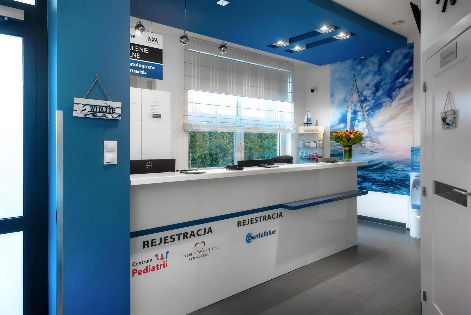
DentalBlue at ul. Kazimierza Pułaskiego 35 has its own radiology unit for dental and panoramic radiographs, CBCT, cephalometric and sinus radiographs. The clinic’s team page lists two people in the role of ‘Radiographer’. Images can be provided on CD with the Romexis viewer; digital images, except intraoral radiographs, are supplied in DICOM format (section 3(4) of the regulation).
The clinic is by the junction with Wspólna, south of the city centre. The ‘Pułaskiego/Wspólna’ stop is 50 m from the entrance and is served by buses 16, 23 and 26. Opening hours are Monday to Friday from 8:00 to 20:00 and Saturday from 8:00 to 16:00; it is closed on Sundays.
Book by calling 85 748 00 50; a receptionist answers during opening hours. Explain which image you need, whether it is your first visit and whether you have a referral. If another practice referred you, ask whether the unit can carry it out. If you are or might be pregnant, mention this when booking. Bring your referral and previous images; Polish, English, Ukrainian and Russian are spoken at the clinic.
Frequently asked questions
How much is a panoramic dental X-ray in Białystok?
DentalBlue’s published price list gives 120 zł for a panoramic radiograph and 60 zł for a sectional panoramic radiograph. A panoramic X-ray is also included in the ‘First visit without pain’ at 300 zł and ‘First visit in pain’ at 340 zł.
Do I need a referral for a panoramic X-ray?
Yes. Panoramic radiographs, cephalometric radiographs and scans are performed on referral or order from a doctor or dentist, even when paid for privately. At a practice with its own radiology unit, the dentist examining you may issue the order.
Does NFZ cover panoramic X-rays in Białystok?
Yes, but only in specified situations alongside treatment with a contracted dentist. Adults can have one for root canal treatment, and any patient before prosthetic treatment, once every 5 years; children aged 5 to 18 can have one in justified cases once every 3 years.
How much does a dental scan cost in Białystok?
DentalBlue lists a CBCT scan of the upper or lower jaw at 300 zł, and both jaws at 350 zł. NFZ does not fund CBCT as part of dental treatment.
Can I have a private tooth X-ray without a referral?
An intraoral radiograph from equipment dedicated solely to these images does not require a referral, but must still be justified, so the dentist decides whether to take it. Panoramic radiographs and scans require a referral or order.
Are dental X-rays safe for a child?
Dental radiograph doses are low, for example below 1.5 µSv for an intraoral image, and ‘Prawo atomowe’ (Atomic Law) provides special protection for people under 16. An image is taken when its result is needed for diagnosis or treatment.
Will I receive the image on disc?
DentalBlue can provide images on CD with the Romexis viewer. Digital images, except intraoral radiographs, are supplied in DICOM format, which allows them to be opened at another practice.
Will DentalBlue take an image on a referral from another practice?
Ask reception on 85 748 00 50 during clinic opening hours. When booking, explain which image the referral specifies.
Sources
- DentalBlue: published price list, X-rays, first visits and RVG items
- Dz.U. 2025 poz. 1288: Rozporządzenie MZ w sprawie warunków bezpiecznego stosowania promieniowania jonizującego dla wszystkich rodzajów ekspozycji medycznej (Minister of Health regulation on safe use of ionising radiation for all types of medical exposure), consolidated text (sections 3 and 5)
- Dz.U. 2026 poz. 1: Prawo atomowe (Atomic Law), consolidated text (Article 3(8a), Articles 33c, 33d and 33e)
- Dz.U. 2025 poz. 615: Rozporządzenie MZ w sprawie świadczeń gwarantowanych z zakresu leczenia stomatologicznego (Minister of Health regulation on guaranteed dental treatment benefits), consolidated text
- Insights into Imaging: Suomalainen A et al., panoramic imaging and CBCT in dental diagnosis, including the European Commission dose table (2015)
- RadiologyInfo (ACR and RSNA): Panoramic Dental X-ray (2024)
An informational article. It does not replace a consultation, and treatment decisions are made during a visit.