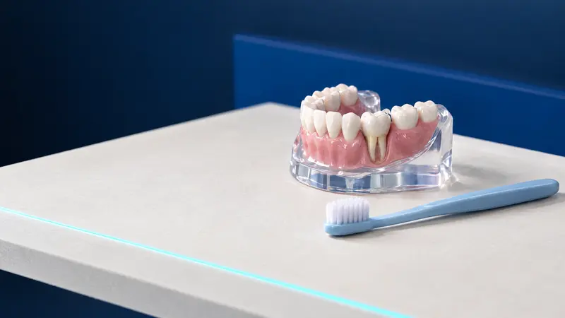
What are cervical tooth lesions, and how do they differ from decay?
Cervical tooth lesions are areas where a tooth has lost enamel and dentine around its neck, at the gum margin, without decay. They are often called wedge-shaped lesions because they commonly form a V-shaped notch, although they can also be shallow, rounded depressions. Nascimento et al.’s review (2016) describes them mainly on the cheek-facing surfaces, more often on incisors and premolars than on canines and molars.
Decay at the tooth neck is caused by plaque bacteria, whereas a non-carious lesion develops without them, through abrasion and acids. However, both processes can affect the same tooth, and decay within a cervical lesion is an indication for a filling. A dental examination therefore establishes the type of lesion.
Cervical tooth lesions are common and become more frequent with age. In a review of 24 studies involving 14 628 people, an average of 46.7% of participants had them: 53% in groups over 30 and 43% in groups under 30 (Teixeira et al., 2020). The table compares the types of lesion.
| Type of lesion | Cause | Typical appearance | What helps |
|---|---|---|---|
| Abrasion | Toothbrush and toothpaste, especially with vigorous scrubbing | Notch at the gum margin, often wedge-shaped | Gentler technique, soft or medium toothbrush, low-abrasion toothpaste |
| Erosion | Acids from food and drinks or the stomach | Shallow, smooth depression with rounded edges | Acidic drinks with meals, reflux treatment, fluoride toothpaste |
| Abfraction, a disputed term | Attributed to excessive biting forces | Sharp V-shaped notch, sometimes on just one tooth | Assess the causes; evidence does not support grinding to adjust the bite |
| Decay at the tooth neck | Plaque bacteria and sugars | Cavity where plaque collects, diagnosed on examination | Decay treatment and a filling, thorough cleaning, fluoride |
What causes wedge-shaped lesions, and is the bite to blame?
Wedge-shaped lesions usually have several causes acting together: Nascimento et al.’s review (2016) stresses that attributing them to a single mechanism is generally a mistake. British Delivering better oral health guidance (DBOH, updated 2025) lists vigorous or inappropriate brushing and highly abrasive toothpastes among the mechanical causes.
Acids come from the diet, for example citrus juices, fizzy drinks and wine, or from the stomach in reflux and eating disorders. The review’s authors therefore recommend a history covering reflux, diet and habits, with medical input when needed.
Abfraction is the most disputed explanation: the theory that biting forces bend the tooth at its neck and cause microcracks in enamel and dentine. The limited clinical studies have not confirmed a clear association between occlusal loading and these lesions, and the review’s authors state that the abfraction theory remains unproven. DBOH recommends avoiding the term altogether.
The practical implication is that grinding to adjust the bite in order to stop cervical lesions has no research support; if done incorrectly, it can increase the risk of decay, wear and sensitivity. There is also insufficient evidence that a night guard prevents abfraction, although the review lists it among ways to limit the effects of grinding.
What symptoms do cervical tooth lesions cause?
Cervical tooth lesions often cause no symptoms and are noticed by a dentist at a check-up. A typical symptom is short, sharp pain with cold or brushing because dentine is exposed in the lesion. DBOH notes that sensitivity can occur with even minor wear, especially at tooth necks, and does not by itself mean progression.
Nascimento et al.’s review (2016) also lists plaque retention within the lesion, changes to the appearance of teeth and weakening of the tooth with deep lesions, which rarely extend as far as the pulp. See a dentist about a lesion in the situations listed below.
- The tooth neck hurts with cold or brushing despite changing your toothpaste and brushing technique.
- The lesion is clearly getting deeper or is visible when you smile.
- Plaque or food collects in the lesion, and its margin extends beneath the gum.
- You have several lesions alongside frequent heartburn, reflux or vomiting.
- Pain lingers after cold or starts without a trigger: dentine hypersensitivity causes brief pain, so these symptoms need an earlier appointment.
Do cervical tooth lesions need fillings?
Not every cervical lesion needs a filling. Nascimento et al.’s review (2016) recommends monitoring symptom-free lesions that do not threaten tooth vitality or function for at least 6 months before planning a procedure. Shallow lesions less than 1 mm deep should be checked every 6 to 12 months, for example at hygiene appointments. Progression is assessed with comparable photographs, models or measurements.
According to the authors, there is no evidence that a filling stops cervical lesions progressing, so fillings are not placed as a precaution; a filling at the tooth neck may also encourage plaque retention. The same review lists situations in which a filling should be considered.
- Decay within the cervical lesion.
- A lesion margin beneath the gum that makes cleaning difficult and increases the risk of decay and periodontal disease.
- Substantial tissue loss that weakens the tooth, or a lesion close to the pulp.
- Persistent sensitivity when approaches without drilling have failed.
- A tooth intended to support a removable denture.
- A patient’s request for cosmetic reasons.
Why does a filling in a cervical lesion fall out?
According to Nascimento et al.’s review (2016), the limited longevity of fillings in cervical lesions remains an unresolved problem. The authors mention difficulty keeping the area dry beside the gum, accessing margins beneath the gum and bonding to sclerotic dentine, the hard, highly mineralised dentine at the base of the lesion. Biting forces and whether the causes have been addressed also matter: fillings fail when scrubbing or acid exposure continues.
The choice of material and bonding system also affects longevity. In a systematic review of clinical studies with at least 18 months of follow-up (Peumans et al., 2014), glass ionomer cements had the lowest average annual rate of filling loss, 2.0%. For composites, the average ranged from 2.5% to 8.4%, depending on the bonding system.
Composite is most often chosen for its appearance. The review’s authors advise using flexible materials that bond well to dentine and resist wear and acids. If a filling at the tooth neck falls out, see a dentist to investigate the cause.
What can you change at home to limit further loss?
Toothpaste cannot rebuild an existing lesion because, according to DBOH, tooth tissue loss is irreversible. You can, however, reduce the factors that enlarge it. DBOH recommends adjusting brushing technique, using a soft or medium toothbrush and standard or low-abrasion toothpaste, and considering fluoride or fluoride-plus-stannous toothpastes for tooth wear.
According to DBOH, there is no strong evidence that brushing timing makes a major difference; vomiting is an exception, after which it is better to rinse the mouth and avoid brushing immediately. The remaining advice is listed below.
- Brush 2 times a day with fluoride toothpaste, without pressing hard or scrubbing.
- Have fizzy and acidic drinks with meals; do not hold them in your mouth or swish them around your teeth.
- Tell your doctor and dentist about frequent heartburn, reflux or vomiting.
- If you clench or grind your teeth, discuss protecting them with your dentist.
- For a dry mouth, chewing sugar-free gum may increase saliva production.
How is sensitivity at the tooth neck treated, and when is a gum graft considered?
Sensitivity in a cervical lesion may ease over time as its dentine gradually mineralises. If it persists, treatment starts with methods that do not involve drilling. Nascimento et al.’s review (2016) lists in-practice fluoride varnish, desensitising agents and a layer of bonding agent, and home products containing potassium nitrate, arginine or high fluoride concentrations. A filling is placed for persistent sensitivity, but does not always help: after 6 months, air or cold sensitivity affected 20 to 30% of restored lesions.
When the lesion is accompanied by gum recession, a graft alone is usually insufficient because the lesion also involves the tooth crown and obscures the enamel-cementum junction. The review describes a combined approach: first a filling to restore tooth shape, then gum repositioning with a connective tissue graft, which systematic reviews report gives the best root-coverage results. Periodontal assessment determines suitability.
How much does treatment cost, and what does the NFZ cover?
The cost depends on whether monitoring is enough or sensitivity treatment, a filling or gum surgery is needed. A cervical lesion usually affects one tooth surface, so a single-surface filling is most commonly involved. The dentist sets the final scope and cost after examination, and prices may change.
Under Poland’s National Health Fund (NFZ), adults are entitled to fillings on 1, 2 or 3 surfaces using glass ionomer cement, including resin-modified cement, and also chemically cured composite in the front teeth from canine to canine. Light-cured composite is available for children and young people under 18 in incisors and canines, and for adults with moderate or severe disability when medically indicated. Fluoride varnishing is included only for those under 18, and recession coverage with a graft is not listed (as of November 2026).
Find practices with an NFZ contract in its ‘Gdzie się leczyć’ (Where to get treatment) search tool. DentalBlue provides treatment according to its published private price list and NFZ-funded treatment under general anaesthesia for patients who meet the criteria.
| Clinic price-list item | Price | When it may be needed |
|---|---|---|
| ‘First visit without pain’ | 300 zł | Examination, panoramic X-ray and written treatment plan |
| ‘Sensitivity treatment, Duraphat, per tooth’ | 40 zł | Fluoride varnish for a sensitive tooth neck |
| ‘Sensitivity treatment, Tokuyama agent’ | 100 zł | Desensitising agent applied in the practice |
| ‘Single-surface filling’ | 300 zł | Restoring a lesion at the tooth neck |
| ‘Occlusal splint’ | 600 zł | Protecting teeth from grinding when recommended by the dentist |
| ‘Coverage of a single gingival recession’ | 1 300 zł | Recession surgery following periodontal assessment |
Cervical tooth lesions at DentalBlue in Białystok
At DentalBlue in Białystok, cervical tooth lesions are assessed during an examination. A ‘First visit without pain’ (300 zł) includes a panoramic X-ray, consultation, examination and written treatment plan; a ‘First visit in pain’ (340 zł) includes a panoramic X-ray, consultation, examination and pain relief. Images are taken in the clinic’s own radiology unit, and its team page lists the role ‘Periodontology specialist’, relevant to gum assessment.
The clinic is at ul. Kazimierza Pułaskiego 35 (15-337 Białystok), at the junction with Wspólna, south of the city centre. The ‘Pułaskiego/Wspólna’ bus stop is 50 m from the entrance, serving routes 16, 23 and 26. Opening hours are Monday to Friday from 8:00 to 20:00 and Saturdays from 8:00 to 16:00; closed Sundays.
Book by phone on 85 748 00 50. Polish, English, Ukrainian and Russian are spoken at the clinic. When booking, explain that you have lesions at the necks of your teeth and say whether cold causes pain. Bring a medicine list, information about reflux, your toothbrush and previous dental images if you have them.
Frequently asked questions
Are cervical tooth lesions dangerous?
Usually not, because tooth wear generally progresses slowly. However, they can cause sensitivity, encourage plaque retention and weaken the tooth when deep, so it is worth having them assessed by a dentist.
Do wedge-shaped lesions need fillings?
Not always. Shallow, symptom-free lesions are usually monitored, while fillings are considered for decay, substantial tissue loss, persistent sensitivity or cosmetic reasons. A filling will not stop further lesions developing if the causes remain.
How much does a filling for a cervical lesion cost?
DentalBlue’s price list shows ‘Single-surface filling’ at 300 zł, ‘Sensitivity treatment, Duraphat, per tooth’ at 40 zł and ‘Sensitivity treatment, Tokuyama agent’ at 100 zł. The dentist sets the scope of treatment and the final cost after the examination, and prices may change.
Can a cervical lesion be filled under the NFZ?
Yes. In a practice with an NFZ contract, adults can receive a glass ionomer cement filling, or chemically cured composite from canine to canine. Light-cured composite is available for adults only with moderate or severe disability and medical indications (as of November 2026).
Is abfraction caused by the bite?
There is no strong evidence for this. Clinical studies have not confirmed a clear link between biting loads and lesions at tooth necks, and DBOH guidance advises against the term abfraction; these lesions are usually associated with several factors together, especially brushing and acids.
Why did a filling at the tooth neck fall out?
It is difficult to keep the area dry at the tooth neck, the lesion margin may be beneath the gum, and hard, sclerotic dentine bonds less well to filling material. Choosing the material and addressing causes such as scrubbing or acids can help.
Sources
- OHID: Delivering better oral health, chapter 7, tooth wear (2021, updated 2025)
- Clinical, Cosmetic and Investigational Dentistry: Nascimento MM et al., abfraction: causes, diagnosis and treatment (2016)
- Journal of Dentistry: Teixeira DNR et al., prevalence of non-carious cervical lesions, review (2020)
- Dental Materials: Peumans M et al., longevity of restorations in non-carious cervical lesions, systematic review (2014)
- ELI: Rozporządzenie MZ w sprawie świadczeń gwarantowanych z zakresu leczenia stomatologicznego (Minister of Health regulation on guaranteed dental services), consolidated text Dz.U. 2025 poz. 615
- DentalBlue: price list
An informational article. It does not replace a consultation, and treatment decisions are made during a visit.