What does a broken instrument in a root canal mean?
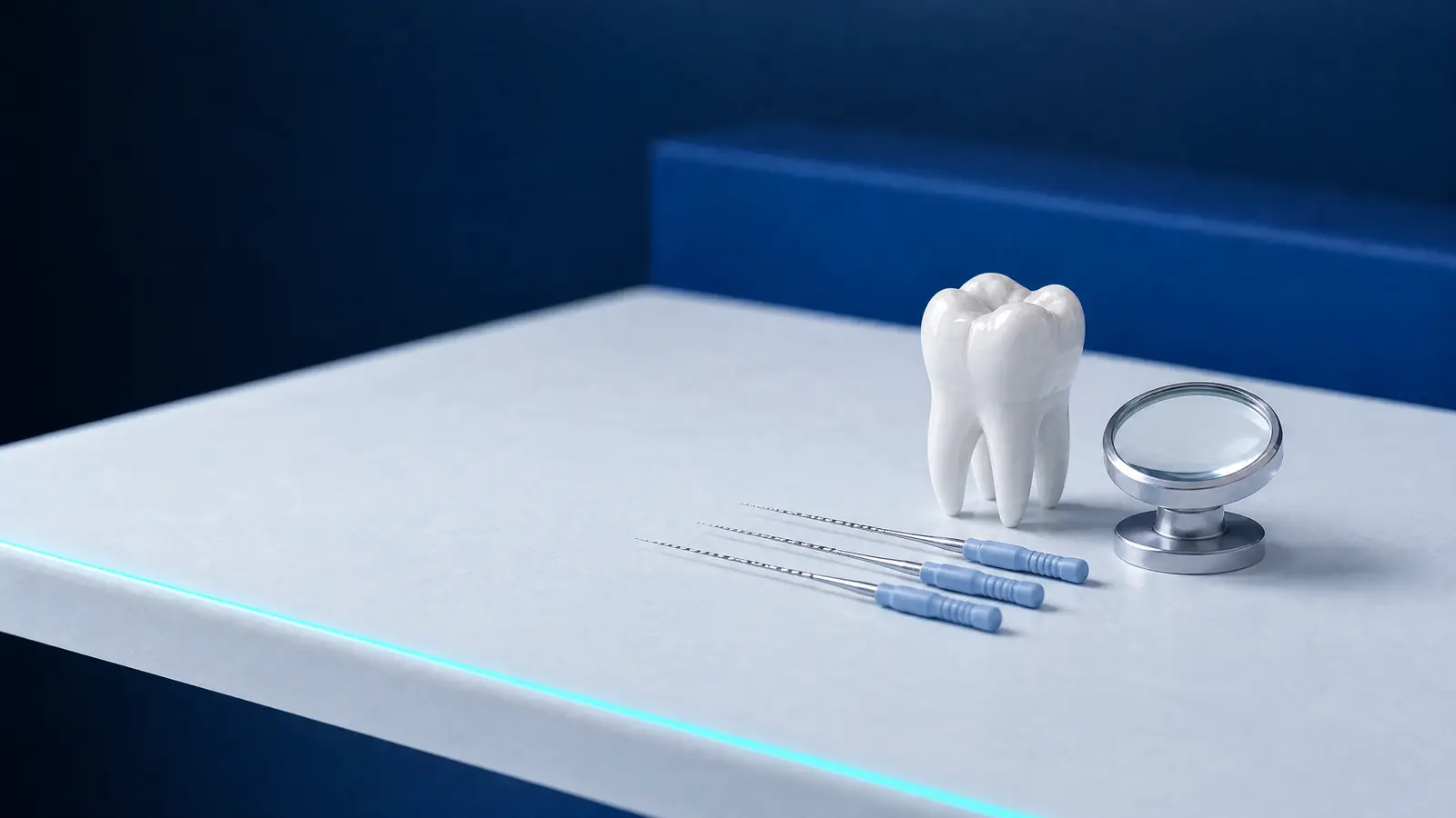
A broken instrument in a root canal is part of an endodontic file, a fine instrument used to clean and widen canals, which separates during use and becomes lodged in the root. Files are made of steel or nickel-titanium and measure fractions of a millimetre across, because they must pass through narrow, often curved canals.
The 2013 review by McGuigan et al. describes two main mechanisms of nickel-titanium instrument fracture: torsional overload and flexural fatigue. Fractures occur mainly in the apical part of the canal; according to this review, their frequency is similar for nickel-titanium and steel instruments.
The fragment stays in the canal, along with whatever lies beyond it. This is why the tooth's prognosis depends less on the piece of metal itself than on whether the infected part of the canal has been cleaned.
How often does a root canal instrument break, and why?
Instrument fracture in a root canal is uncommon. An analysis of records for 8 460 cases from an Australian specialist practice found a retained instrument fragment in 3.3% of treated teeth (Spili et al., 2005); a 2017 Swiss review calls instrument fracture a rare adverse event.
Risk depends primarily on tooth anatomy. In a series of 97 instrument-fracture cases, instruments broke more often in curved canals than straight ones, and half the fractures involved the mesial roots of lower molars (Suter et al., 2005).
McGuigan et al. identify operator skill, manufacturers' improvements to alloys and limiting instrument reuse as factors reducing fracture frequency. Fracture is therefore a recognised risk of root canal treatment that cannot be eliminated completely.
Is a broken instrument in a root canal dangerous?
A broken instrument in a root canal is usually not dangerous in itself, and the tooth's prognosis often remains similar to that without a fragment. In a comparison of 146 teeth with retained fragments and 146 matched control teeth, healing was 91.8% and 94.5%, with no statistically significant difference (Spili et al., 2005).
A 2010 meta-analysis reported average healing of 91% in teeth with retained fragments and concluded that the fragment itself did not significantly worsen prognosis (Panitvisai et al., 2010). An inflammatory lesion around the root did make a difference: 80.7% of lesions present before treatment healed, whereas 92.4% of teeth without lesions remained healthy.
The 2013 review by McGuigan et al. concludes that a fragment does not worsen prognosis when there is no inflammation around the root, but healing is clearly poorer when a lesion already exists. Timing therefore matters: the earlier a file breaks during canal cleaning, the more bacteria may remain beyond it.
What are the options: removal, bypassing, leaving it or surgery?
When an instrument breaks in a canal, the dentist has several options: remove the fragment, bypass it, leave it and monitor, or operate. The 2013 review by McGuigan et al. recommends choosing according to the prognosis of each option, balanced against the risks of removing or retaining the fragment.
The table outlines these approaches. It is a general framework from the literature; for an individual tooth, the dentist decides after an X-ray, often a CT scan and examination under a microscope.
| Option | What it involves | When it is considered |
|---|---|---|
| Removal under a microscope | Exposing the fragment, loosening it with ultrasound and retrieving it | Accessible fragment in the coronal or middle part, a lesion around the root |
| Bypassing the fragment | Passing a fine instrument beside the fragment and preparing the canal | Space between the fragment and canal wall |
| Leaving it and monitoring | Filling the canal up to the fragment and follow-up X-rays | No inflammatory lesion, fragment deep in a curved canal |
| Apicectomy | Surgically removing the root tip with the fragment | Fragment near the tip, a lesion that does not heal after treatment |
| Tooth extraction | Extraction and planning replacement of the missing tooth | A tooth that cannot be treated further or restored |
How is a broken root canal instrument removed, and how often does it work?
A broken instrument is removed under a microscope: the dentist creates straight-line access to it, exposes its upper end with ultrasonic tips, vibrates it and flushes it out or retrieves it using special techniques. In the series of 97 cases by Suter et al. (2005), removal succeeded in 84, or 87%; the authors considered an operating microscope essential for these techniques.
Success depends on fragment position. A 2024 review of 11 studies covered 1 133 treated teeth: removal failed in 172, and perforation, or breaching the root wall, occurred in 55; failure rates were 21% for fragments in the apical part and 8.8% in the middle or coronal part (Dioguardi et al., 2024).
Removal also has a biological cost. In the study by Souter and Messer (2005), perforations and failed attempts occurred only with apical fragments, and root strength decreased more with deeper fragments. Suter et al. noted that success fell as the procedure took longer, so the dentist judges when further attempts are no longer worthwhile.
When is it better to leave a fragment in the canal?
Leaving a fragment in the canal is preferable when there is no inflammatory lesion around the root and removal would considerably weaken it. McGuigan et al. (2013) write that removal may be unnecessary in these cases and retaining or bypassing the fragment should be considered, because removal carries significant risks, especially in the apical part.
Souter and Messer (2005) advise against routinely attempting to remove fragments from the apical part of curved canals. The canal is then filled with a tight seal up to the fragment, and the tooth restored as after any root canal treatment.
A retained fragment requires follow-up. European Society of Endodontology (ESE) guidelines recommend assessing the outcome of root canal treatment after at least one year, with further reviews until healing or for 4 years if a lesion around the root persists; apicectomy is considered if the lesion does not heal.
What you should know as a patient: information and records
As a patient, you have a right to know that an instrument fragment remains in your tooth's canal. The Polish Act on Patients' Rights and the Patient Rights Ombudsman (Rzecznik Praw Pacjenta; t.j. Dz.U. 2024 poz. 581), in art. 9 ust. 2, grants a right to accessible information about diagnosis, proposed and possible treatments, their foreseeable consequences, treatment outcomes and prognosis (legal status as of 24 October 2026).
McGuigan et al. (2013) stress that the patient should be informed and the event recorded, whether the instrument breaks during treatment or the fragment is noticed on a routine X-ray. The 2006 ESE guidelines also recommend documenting root canal complications, including iatrogenic events.
Art. 23 ust. 1 of the same Act grants access to medical records, and art. 28 ust. 2a provides for the first copy or printout within the requested scope to be supplied free of charge. Previous X-rays help the next dentist assess fragment position, and the ESE considers it appropriate to obtain images from the previous dentist, especially for a tooth that has already had root canal treatment.
How much does removing a broken root canal instrument cost?
The cost of removing a broken instrument depends on the number of visits and whether the attempt succeeds. In the clinic's price list, “Usunięcie złamanego narzędzia pod mikroskopem jedna wizyta i każda” (broken instrument removal under a microscope, each visit) costs 250 zł, “Usunięcie złamanego narzędzia kanałowego z innego gabinetu” (removal of a root canal instrument broken at another practice) 350 zł and “Niepowodzenie w usunięciu złamanego narzędzia kanałowego” (unsuccessful broken root canal instrument removal) 200 zł.
Root canal treatment or retreatment itself, priced by the number of canals, and restoration of the tooth are additional costs; the articles on root canal costs and retreatment explain the details. The dentist sets the final scope and cost after examination, and prices may change.
How it works at DentalBlue
At DentalBlue in Białystok, root canal treatment and retreatment are also carried out under a microscope, and the price list includes broken instrument removal and perforation repair. The clinic's own radiology unit takes intraoral and panoramic X-rays and CBCT scans, so fragment position can be assessed on site. Bring records and X-rays from previous treatment to the consultation. The price list item “Konsultacja przy użyciu mikroskopu” (consultation using a microscope) costs 300 zł; the dentist determines the final scope and cost, and prices may change.
Frequently asked questions
Can a broken file in a root canal cause infection?
The fragment itself is not usually the cause of infection. Bacteria in the part of the canal beyond it that could not be cleaned may be the problem. The 2017 Swiss review stresses that outcome depends on when the fracture occurred and the degree of canal infection, rather than the fragment's mere presence.
Can a broken instrument be seen on an X-ray?
Yes, the metal fragment is visible on an X-ray, so it is sometimes discovered incidentally, even years later. McGuigan et al. (2013) stress that patients should also be informed when a fragment is noticed on a routine X-ray, and this must be documented.
Must an instrument be removed if the tooth does not hurt?
Not always. When a tooth does not hurt and there is no lesion around the root on the X-ray, McGuigan et al. (2013) recommend considering retaining or bypassing the fragment, because removal carries risks. The tooth is monitored with X-rays, with treatment considered if a lesion appears.
How many visits does broken instrument removal take?
It depends on the fragment's position and length and the canal's curvature: sometimes one visit is enough, sometimes more are needed. In Suter et al. (2005), success fell as the procedure took longer, so the dentist assesses when to stop attempts and choose another approach.
Can I continue treatment at another practice with an instrument fragment in the canal?
Yes. You have a right to access your medical records, and the first copy within the requested scope is free (art. 23 and 28 of the Polish Act on Patients' Rights). X-rays and details of previous treatment help the next dentist plan management.
Sources
- Journal of Endodontics: Spili P, Parashos P, Messer HH, instrument fracture and root canal treatment outcome (2005)
- Journal of Endodontics: Panitvisai P et al., retained instrument fragments and treatment outcome, meta-analysis (2010)
- British Dental Journal: McGuigan MB et al., clinical decisions after endodontic instrument fracture (2013)
- International Endodontic Journal: Suter B et al., probability of removing broken instruments from canals (2005)
- Healthcare: Dioguardi M et al., broken instrument removal during retreatment, meta-analysis (2024)
- Journal of Endodontics: Souter NJ, Messer HH, complications of ultrasonic broken instrument removal (2005)
- Swiss Dental Journal: Suter B, fractured root canal instruments, incidence, location and treatment (2017)
- ISAP: Ustawa o prawach pacjenta i Rzeczniku Praw Pacjenta, t.j. Dz.U. 2024 poz. 581
An informational article. It does not replace a consultation, and treatment decisions are made during a visit.