Where do white spots on teeth come from?
White spots on teeth have two main groups of causes: demineralisation of the enamel by dental plaque, which appears on a tooth already in the mouth, and disorders of enamel development that arose before the tooth came through. The first group includes early decay and spots after fixed braces, the second MIH (molar incisor hypomineralisation) and fluorosis.
The distinction matters in practice, because demineralisation can deepen or stop, while a developmental spot has been there since the tooth erupted and does not disappear with better brushing. An infection in a baby tooth can also harm the permanent tooth developing beneath it, but there is little research on this. The table sets out the most common causes.
| Cause | What it usually looks like | Who it affects | What is usually done |
|---|---|---|---|
| Demineralisation, early decay | A matt white spot next to the gum or between teeth | Any age, where plaque builds up | Oral hygiene, fluoride, sometimes resin infiltration |
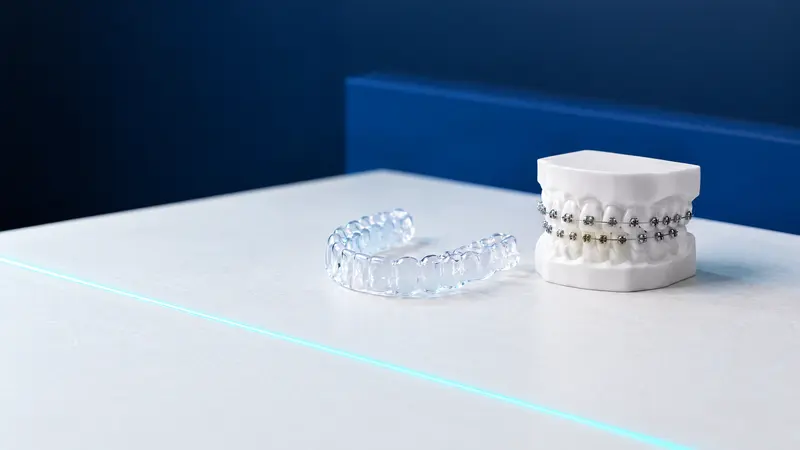
| Spots after fixed braces | White patches around where the brackets were | Up to about half of people treated with fixed braces | Monitoring, fluoride, possibly infiltration later |
| MIH (molar incisor hypomineralisation) | Clearly demarcated spots on the first molars, often also on the incisors | About 13 to 14% of children worldwide | Prevention, fillings, crowns, extractions in severe forms |
| Fluorosis | Fine white lines or flecks, pitting in more severe forms | Excess fluoride swallowed while the teeth are developing | Masking with infiltration, veneers for extensive changes |
Are white spots on teeth a sign of decay?
A white spot on a tooth can be the earliest stage of decay, before a cavity forms. The American Academy of Pediatric Dentistry (AAPD) counts such lesions without a cavity as early childhood caries, so a white spot next to the gum in a young child is a reason for a visit, not just for watching at home.
Demineralisation without a cavity can often be stopped. A 2015 Cochrane review showed that sealing or resin infiltration reduced the chance of early lesions on the contact surfaces between teeth progressing (OR 0.24) compared with fluoride varnish or advice to floss alone. In a 3-year study by Meyer-Lückel, progression occurred in 4% of lesions after infiltration and in 42% of control lesions.
Developmental spots, such as MIH and fluorosis, are not decay, but enamel affected by MIH is often weaker and crumbles more easily. Whether a given spot is demineralisation or a developmental defect is assessed by the dentist during an examination, sometimes with the help of an X-ray.
White spots after braces: why do they appear and do they go away?
White spots after fixed braces are areas of demineralised enamel around the brackets, where plaque collects and is harder for a toothbrush to reach. A review by Sundararaj et al. (2015) reports that new spots appear during treatment with fixed braces in up to about half of patients, with wide variation between studies.
Some spots fade on their own after the braces come off. In a study by Sonesson et al. (2021), the number of people with spots fell by more than half within a year of the braces being removed, both with fluoride varnish and with placebo. That is why it is usually worth waiting and assessing what remains before deciding on treatment.
Preventing the spots is better, though. A 2019 Cochrane review found low-certainty evidence that a 12 300 ppm fluoride foam used every 6 to 8 weeks and a 5000 ppm toothpaste reduce the number of new spots during treatment. A 2026 meta-analysis indicates that no product significantly reduced the size of spots that had already formed.
Molar incisor hypomineralisation (MIH): what is it and how is it treated?
Molar incisor hypomineralisation (MIH) is a clearly demarcated defect in the mineralisation of enamel that affects one to four of the first permanent molars, often together with the incisors. A meta-analysis by Schwendicke et al. (2018) estimates that it affects 13.1% of children worldwide, and regional Polish studies report from about 6 to 14%.
The causes of MIH are multifactorial. In its 2022 recommendations, the European Academy of Paediatric Dentistry (EAPD) lists lack of oxygen and other problems around birth, premature birth, illnesses in infancy and early childhood, and genetic predisposition. It is not the parents’ fault, nor the result of poor brushing.
A tooth with MIH is often sensitive, and the enamel may crumble: according to the same meta-analysis, about 27.4% of cases need treatment because of pain, sensitivity or enamel breakdown. The EAPD states that in molars, composite fillings, preformed metal crowns and laboratory-made restorations are highly effective, and in severe cases a planned extraction of the tooth is considered. The choice depends on the severity of the changes and the child’s age.
Dental fluorosis: where does it come from, and is fluoride toothpaste safe?
Fluorosis is an enamel disorder caused by swallowing too much fluoride during the period when the teeth are developing in the bone. It ranges from fine white lines and flecks to pitting, which may in time take on brown stains. For the permanent incisors, the key age is about 20 to 30 months.
Fluorosis is not a reason to give up fluoride toothpaste, only to use an amount suited to the child’s age. The 2019 EAPD recommendations provide for a 1000 ppm toothpaste in a rice-grain-sized smear from the first tooth to the age of 2, and a pea-sized amount from 2 to 6 years, and from the age of 6 a 1450 ppm toothpaste. Until at least the age of 7, a parent supervises toothbrushing.
In mild fluorosis, treatment is usually aesthetic. When the spots are bothersome, they can be masked with resin infiltration, and for extensive changes the dentist may suggest veneers or bonding. In studies of fluorosis, whitening before infiltration did not improve the masking effect.
Resin infiltration (ICON): what does it achieve and what can it not promise?
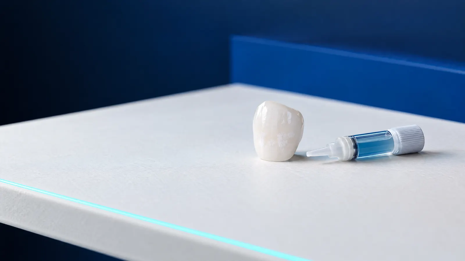
Resin infiltration is a micro-invasive method: porous, demineralised enamel is saturated with a liquid resin, without drilling. The procedure has two aims: to stop early decay from progressing and to mask the white spot so that it stands out less from the rest of the tooth.
For masking, the results are promising but mixed. A review by Bourouni et al. (2021) found a better optical effect from infiltration than from no treatment and than from fluoride varnish (moderate-quality evidence). A more recent meta-analysis by Ståhl et al. (2026), however, describes the results as highly inconsistent: patient satisfaction rose after one month, but after 12 months the differences in the colour and size of the spots were no longer significant.
Infiltration can therefore improve how the spots look, but it does not always make them disappear, and the effect varies from person to person. That is why, before the procedure, the dentist assesses whether the spot is demineralisation or a developmental defect and discusses realistic expectations.
What can you do at home, and when should a white spot be seen by a dentist?
At home, white spots are best protected in the same way as the whole tooth: by brushing thoroughly twice a day with fluoride toothpaste and cutting down on frequent snacking on sweets. The UK guidance “Delivering better oral health” advises adults to use a toothpaste with 1350 to 1500 ppm fluoride and to spit out the foam after brushing without rinsing with water. It is worth booking a visit if any of the signs on the list appear; more on how children should brush their teeth can be found in the article on tooth decay in children.
- A new white spot in a child, especially along the gum line.
- A spot that grows, becomes rough, darkens or turns into a cavity.
- Crumbling enamel, or pain when brushing and eating, in a child’s newly erupted molars.
- Spots around the brackets during treatment with fixed braces.
- Spots that are an aesthetic concern and whose cause is not known.
How it works at DentalBlue
At DentalBlue in Białystok, the cause of white spots is assessed by a dentist at a visit without pain, which includes a panoramic X-ray, a consultation, an examination and a written treatment plan; in the clinic’s price list it costs 300 zł. The item “icon-infiltracja żywicą” (Icon resin infiltration) costs 350 zł, “Fluoryzacja” (fluoride treatment) 100 zł, and “Bonding 1 zęba” (bonding, one tooth) 1 000 zł. The final scope and cost are set at the consultation, and prices may change. The clinic also provides paediatric dentistry and orthodontics, so spots in children and during treatment with braces are assessed in the same place.
Frequently asked questions
Can white spots on teeth be whitened?
Whitening lightens the whole tooth but does not remove the spot itself, and in studies of fluorosis, whitening before infiltration did not improve the masking effect. The dentist chooses the method after establishing whether the spot is demineralisation or a developmental defect.
Are white spots on a child’s teeth caused by a lack of calcium?
The cause is rarely that simple. For MIH, the EAPD points to problems around birth, premature birth, illnesses in early childhood and genetics; for fluorosis, to excess fluoride; and for demineralisation, to dental plaque. It is not worth giving supplements on your own initiative; the cause is established by the dentist.
Will white spots after braces go away on their own?
Some will. In the study by Sonesson et al., the number of people with spots fell by more than half within a year of the braces being removed. That is why a decision on infiltration is usually made after a few months of monitoring, with careful oral hygiene and fluoride toothpaste.
Does a tooth with MIH decay faster?
It can, because enamel affected by MIH is often weaker, crumbles and is sensitive, which makes brushing harder. A 2018 meta-analysis reports that about 27.4% of cases need treatment. More frequent check-ups and fluoride help to catch changes early.
Are white spots on baby teeth a sign of decay?
They may be the start of decay, especially matt spots along the gum line. The AAPD counts such lesions without a cavity as early childhood caries. At this stage the process can often be stopped with oral hygiene and fluoride, so it is worth showing them to a dentist.
Sources
- Journal of Dentistry: Schwendicke F et al., global prevalence of MIH, meta-analysis (2018)
- European Archives of Paediatric Dentistry: Lygidakis NA et al., EAPD recommendations on MIH (2022)
- Advances in Clinical and Experimental Medicine: Głódkowska N, Emerich K, MIH in Silesia and Pomerania (2020)
- European Journal of Orthodontics: Sonesson M et al., white spots after removal of braces (2021)
- Cochrane: Benson PE et al., preventing white spots during treatment with fixed braces (2019)
- Clinical Oral Investigations: Bourouni S et al., resin infiltration of white spots and fluorosis (2021)
- European Journal of Orthodontics: Ståhl L et al., results of resin infiltration over time (2026)
- Cochrane: Dorri M et al., micro-invasive treatment of early caries (2015)
An informational article. It does not replace a consultation, and treatment decisions are made during a visit.