What is the periodontium and what does it consist of?
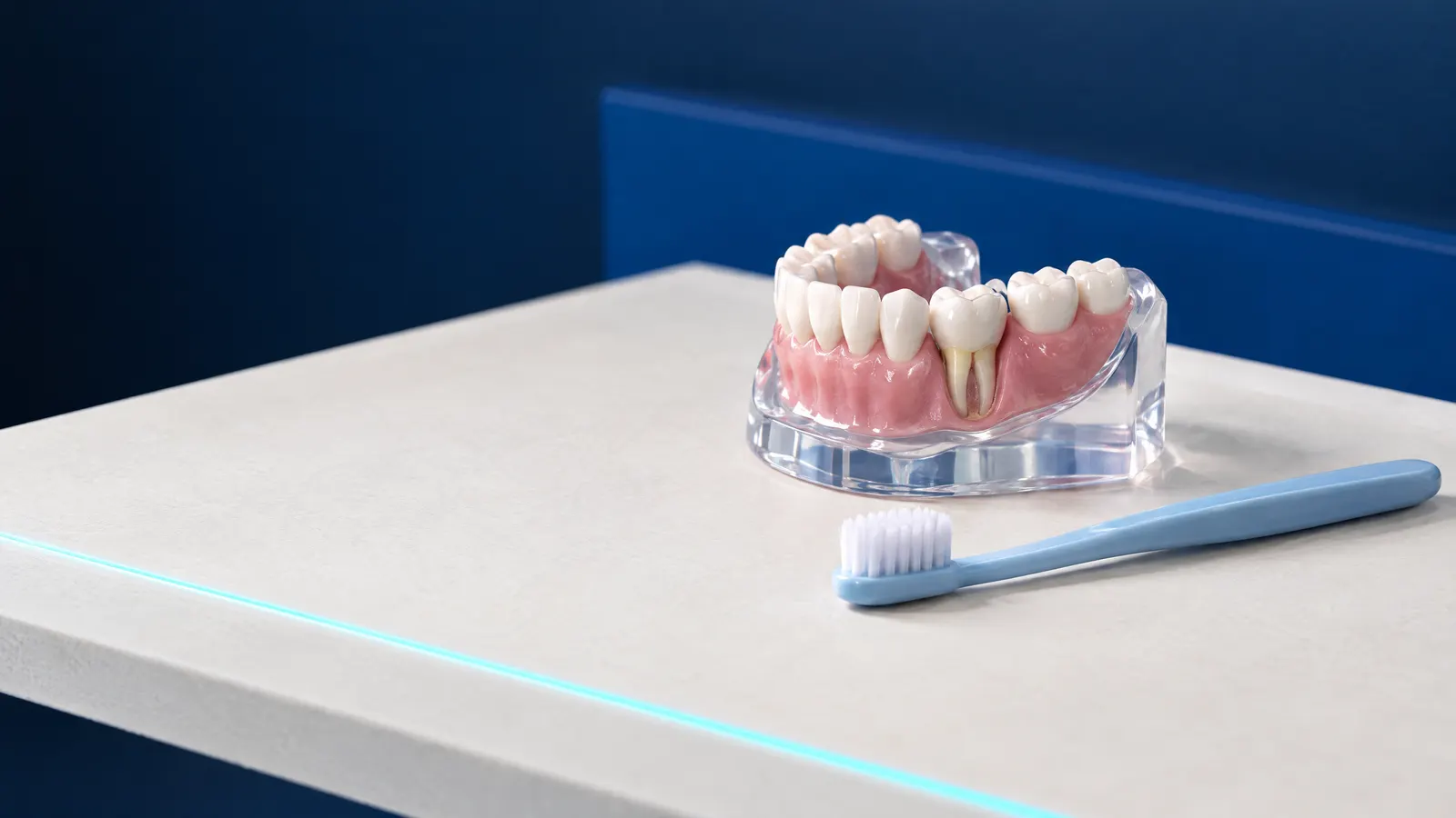
The periodontium is the group of tissues surrounding a tooth that hold it in the upper or lower jawbone. It has four main components: the gum, the periodontal ligament connecting the root to the bone, root cementum covering the root, and alveolar bone. The Medycyna Praktyczna medical portal also includes the alveolar mucosa among the periodontal tissues.
The technical term is periodontium, which gives the specialty of periodontology its name. Periodontal diseases usually start in the gum, but cause their most serious effects when they destroy the ligament and bone: better brushing alone cannot reverse that loss. The table shows the periodontal tissues and their functions.
| Part of the periodontium | What it is | Its function |
|---|---|---|
| Gum | Mucous membrane over the alveolar process, around the necks of the teeth | Together with periodontal ligament fibres, forms a barrier between the root and bacteria in the mouth |
| Periodontal ligament | Collagen fibres, blood vessels and nerves between the root and bone | Suspends the tooth in its socket, cushions chewing forces and transmits sensations of touch, pressure and pain |
| Root cementum | A thin mineralised layer on the root | Anchors periodontal ligament fibres on the tooth side |
| Alveolar bone | The part of the upper and lower jaws containing the tooth sockets | Supports the tooth and anchors the periodontal ligament; vessels supplying the ligament pass through it; it is constantly remodelled |
How does the periodontium work, and why does a healthy tooth move slightly?
A tooth is not fused to the bone: it is suspended in its socket by bundles of collagen fibres, the periodontal ligament. Even a healthy tooth therefore has physiological mobility of 0.08 to 0.15 mm, invisible to the eye. Fibres near the tooth neck stabilise it sideways and separate the socket from the mouth, while deeper fibres resist the tooth being pushed into the bone during chewing.
The periodontal ligament contains nerves that transmit touch, pressure and pain, and its cells contribute to healing and remodelling of bone, cementum and fibres. When periodontitis destroys the ligament and bone, the tooth loses support and may become noticeably loose; tooth mobility is covered in a separate article.
What does a healthy periodontium look like?
A healthy periodontium has no clinically detectable inflammation. Under the 2017 classification of periodontal diseases, developed by the American Academy of Periodontology (AAP) and the European Federation of Periodontology (EFP), fewer than 10% of sites bleed on gentle probing, and there has been no loss of attachment or bone due to previous periodontitis.
A reduced periodontium, with some attachment already lost, can also be healthy, for example after successful periodontitis treatment. The 2020 EFP guideline considers a treated patient stable when fewer than 10% of sites bleed, pockets measure up to 4 mm, and no 4 mm site bleeds.
However, as the classification authors emphasise, such a person remains a periodontitis patient for life and needs supportive care because recurrence is possible. Gingivitis, by contrast, can resolve completely. Show your dentist any symptoms on the list.
- Gums bleeding when brushing or flossing.
- Red or swollen gums.
- Receding gums and exposed roots.
- Loose teeth or new gaps between them.
- Swelling with pus beside a tooth: call the dental practice the same day.
How dentists examine the periodontium: probing, bleeding and X-rays
Periodontal examination centres on probing: the dentist gently inserts a thin, graduated probe between the gum and tooth, measures the pocket depth and checks for bleeding. In the 2017 classification, bleeding on probing is the main parameter distinguishing healthy gums from inflammation.
The British Society of Periodontology’s BPE (Basic Periodontal Examination) is a quick screening examination; a similar index is sometimes called PSR. The highest score is recorded in each of 6 sextants using a WHO probe with a black band from 3.5 to 5.5 mm. BPE is for screening only, not diagnosis.
For code 4, the guidance recommends recording pocket depths and bleeding at 6 points around every tooth; for code 3, this charting is carried out in the affected sextant after initial treatment. X-rays showing bone levels are also needed, with the dentist choosing the type. BPE is not used around implants.
- Code 0: pockets shallower than 3.5 mm, with no calculus or bleeding.
- Code 1: as above, but with bleeding; oral hygiene instruction is sufficient.
- Code 2: calculus or an overhanging filling margin; deposit removal is also needed.
- Code 3: a pocket from 3.5 to 5.5 mm, usually 4 to 5 mm.
- Code 4: a pocket deeper than 5.5 mm; specialist care is sometimes needed.
- An asterisk beside the code indicates involvement of the root furcation.
Periodontal diseases: what does the 2017 classification say?
The current classification of periodontal and peri-implant diseases was adopted at the AAP and EFP workshop in Chicago in November 2017. It covers the four main groups listed below.
Gingivitis is diagnosed when at least 10% of sites bleed and there is no attachment loss: 10 to 30% indicates localised disease, and above 30% generalised disease. Periodontitis is diagnosed by the dentist from a characteristic pattern of attachment loss and bone assessment, after other causes of tissue damage have been ruled out; one criterion is interdental attachment loss at two or more teeth that are not next to each other.
The classification describes recession through interdental attachment loss, tissue thickness and the appearance of the exposed root. Around implants, mucositis causes bleeding on probing and resolves after plaque removal, while peri-implantitis also involves progressive bone loss and is more common with poor plaque control and a history of severe periodontitis.
- Periodontal health and gingival diseases, whether plaque-induced or unrelated to plaque.
- Periodontitis, commonly called gum disease, described by stage and grade, alongside necrotising forms and periodontitis as a manifestation of systemic disease.
- Other conditions affecting the periodontium, including gum recession and occlusal trauma.
- Peri-implant conditions: health, mucositis and peri-implantitis.
What do stages I to IV and grades A, B and C mean?
Every case of periodontitis is described by two features. The stage, from I to IV, indicates disease severity and treatment complexity and, under the EFP guideline, forms the basis of the treatment plan. The grade, from A to C, describes the rate of progression and risk of further disease; the dentist initially assumes grade B and changes it when there is evidence to do so.
Grade C means rapid progression, for example loss of at least 2 mm of attachment or bone over 5 years. Risk factors modify the grade: smoking fewer than 10 cigarettes a day corresponds to grade B, and 10 or more to grade C; diabetes with HbA1c below 7% corresponds to grade B, and 7% or above to grade C.
Extent is added to the stage: localised disease affects fewer than 30% of teeth. The stage does not decrease after treatment. The table shows simplified criteria.
| Stage | Interdental attachment loss | Bone loss on X-ray | Typical features |
|---|---|---|---|
| I | 1 to 2 mm | Less than 15% of root length | Pockets up to 4 mm, mainly horizontal bone loss |
| II | 3 to 4 mm | 15 to 33% of root length | Pockets up to 5 mm, mainly horizontal bone loss |
| III | 5 mm or more | Extending to the middle or apical part of the root | Pockets of 6 mm or more, vertical bone defects of 3 mm or more, up to 4 teeth lost due to disease |
| IV | 5 mm or more | Extending to the middle or apical part of the root | At least 5 teeth lost because of periodontitis, or complex problems needing rehabilitation, such as bite collapse or marked tooth mobility; not all features have to be present at once |
What harms the periodontium? Main risk factors
The EFP guideline describes periodontitis as a chronic inflammatory disease with multiple causes, associated with a disturbed bacterial biofilm: plaque at the gum line and between teeth. The factors listed affect the disease course and response to treatment.
Excessive bite forces are often said to destroy the periodontium. However, the classification authors state that human studies linking occlusal trauma to progressive attachment loss in periodontitis are lacking.
- Smoking: raises the disease grade, and the EFP includes support with quitting in the first treatment step.
- Diabetes, particularly when poorly controlled: HbA1c of 7% or above indicates grade C, and better diabetes control is also part of the first step.
- Calculus and overhanging filling margins that retain plaque.
- Previous periodontitis, which increases the risk of recurrence and, around implants, peri-implantitis.
Gum specialists: who treats the periodontium and what does the NFZ cover?
A specialist in gums and periodontal tissues is a periodontist, a dentist specialising in periodontology. A general dentist can carry out screening and remove calculus; the BPE guidance recommends considering specialist referral for pockets deeper than 5.5 mm.
Under Poland’s National Health Fund (NFZ), the oral surgery and periodontology benefits list is provided by, among others, periodontology specialists and dentists training in that specialty; contracted clinics appear in the NFZ ‘Gdzie się leczyć’ (Where to get treatment) search tool. NFZ-funded open curettage is available only to people under 18 and women during pregnancy and the postpartum period; regenerative procedures and recession coverage are not included in the lists. Adults are entitled to the listed services (as at November 2026).
- An examination with oral hygiene instruction once per calendar year.
- Calculus removal from all teeth once per calendar year.
- Pocket irrigation with medication application and treatment of mucosal lesions, billed per visit.
- Closed curettage within one quadrant.
- A specialist consultation with a written assessment for the treating dentist.
- A panoramic X-ray for suspected periodontitis, once every 3 years, on referral from a specialist or a dentist in specialist training.
Periodontal examination at DentalBlue in Białystok
At DentalBlue in Białystok, periodontal assessment and treatment form part of periodontology and preventive care, from consultations and hygiene treatment to surgery. The clinic’s team page lists the roles ‘Periodontology specialist’ and ‘Head of dental hygienists’. Panoramic and intraoral X-rays and, where indicated, CBCT scans are carried out in the clinic’s own radiology unit.
The table lists items from the published private price list. Closed curettage is not listed separately, so the dentist gives its cost in the treatment plan; prices may change, and the final scope and cost are agreed at consultation.
The clinic is at ul. Kazimierza Pułaskiego 35, near the junction with Wspólna, south of the city centre; the ‘Pułaskiego/Wspólna’ stop is 50 m from the entrance and served by buses 16, 23 and 26. Opening hours are Monday to Friday 8:00 to 20:00 and Saturday 8:00 to 16:00; the clinic is closed on Sundays.
Book by calling 85 748 00 50 during opening hours. Say you need a gum and periodontal assessment and describe symptoms such as bleeding or receding gums. You can speak Polish, English, Ukrainian or Russian.
- Bring a medication list and information about long-term conditions, especially diabetes, with your HbA1c result if available.
- Bring previous dental images and pocket measurement records.
- Tell the dentist whether you smoke and how much, as this affects the disease grade.
| Price-list item | Price | Purpose |
|---|---|---|
| ‘Periodontal consultation’ | 300 zł | Periodontal examination and treatment plan |
| ‘First visit without pain’ | 300 zł | Includes a panoramic X-ray and written treatment plan |
| ‘Complete hygiene treatment (scaling, air polishing, fluoride)’ | 350 zł | Removal of calculus and deposits |
| ‘Periodontal pocket irrigation’ | 100 zł | As indicated by the dentist |
| ‘Open curettage, one tooth’ | 400 zł | One tooth with a deep pocket |
| ‘Panoramic radiograph’ | 120 zł | Assessment of bone levels |
Frequently asked questions
How is the periodontium different from the gum?
The gum is just one visible part of the periodontium. The periodontium also includes hidden tissues: the periodontal ligament, root cementum and alveolar bone, which is why gingivitis and periodontitis are two different diseases.
Can the periodontium regenerate?
Inflamed gums return to health once plaque and calculus are removed. Lost bone does not grow back on its own; for vertical bone defects of 3 mm or more, the EFP guideline recommends regenerative surgery, but the main aim of treatment is to stop the disease.
How can I tell whether I have periodontal disease?
Bleeding gums, recession or loose teeth can be warning signs. Only an examination can confirm it: probing pocket depths with a bleeding assessment and an X-ray showing bone levels.
How much does a periodontal examination cost?
DentalBlue’s published price list gives a ‘Periodontal consultation’ at 300 zł. A ‘First visit without pain’ costs the same and includes a panoramic X-ray, consultation, examination and written treatment plan; prices may change.
Does the NFZ cover periodontal examination?
Yes. For adults, the NFZ funds an examination with hygiene instruction once a year, an oral surgery and periodontology specialist consultation and, for suspected periodontitis, a panoramic X-ray once every 3 years on specialist referral (as at November 2026).
Sources
- Medycyna Praktyczna (mp.pl): Tomaszewska IM, structure of teeth and the periodontium (2016)
- Journal of Clinical Periodontology: Caton JG et al., new classification of periodontal and peri-implant diseases, introduction (2018)
- Journal of Clinical Periodontology: Sanz M et al., EFP S3 guideline on treatment of stage I to III periodontitis, including staging and grading criteria (2020)
- British Society of Periodontology: Basic Periodontal Examination (BPE), guidelines (2019)
- ELI: Rozporządzenie MZ w sprawie świadczeń gwarantowanych z zakresu leczenia stomatologicznego (Health Minister’s regulation on guaranteed dental benefits), consolidated text Dz.U. 2025 poz. 615 (Annexes 1 and 5)
- DentalBlue: published price list (periodontology, hygiene, first visit, radiology)
An informational article. It does not replace a consultation, and treatment decisions are made during a visit.