What do doctors mean by oral infection foci?
Oral infection foci, also called sources of odontogenic infection, are sites of chronic infection in teeth, periodontal tissues or jawbones from which bacteria or inflammatory products may affect the rest of the body. In a 2000 review, Pallasch and Wahl define focal infection as local or systemic infection caused by microorganisms or toxins spreading from a single focus.
In practice, this includes several types of lesion a dentist can find by examination and imaging. The list covers those considered when another specialist asks for dental infection foci to be excluded.
- A dead tooth with an inflammatory root-tip lesion: a granuloma, cyst or chronic abscess.
- A root-treated tooth with symptoms or a root lesion that the dentist, after comparing X-rays, judges to be persistent disease rather than healing.
- Deep decay reaching the pulp and causing inflammation.
- Periodontitis with deep pockets and periodontal abscesses.
- A partly erupted or impacted wisdom tooth with recurrent inflammation of the surrounding gum.
- Infected roots left in the bone after a tooth has fractured or broken down.
Why do infection foci often cause no pain?
Infection foci often cause no pain because infection around the root or in periodontal tissues develops slowly and chronically. The American Association of Endodontists (AAE) describes asymptomatic apical periodontitis as a root-tip lesion visible on an X-ray without clinical symptoms.
A 2021 review by Tibúrcio-Machado et al., covering 114 studies and over 34 000 people, reports lesions in 52% of participants, at 5% of all teeth and 39% of previously root-treated teeth. The authors stress that X-rays are needed for diagnosis because lesions are often symptom-free.
In a histological study of 256 root lesions from extracted teeth, Nair et al. (1996) found 50% granulomas, 35% abscesses and 15% cysts. The article on dental cysts explains how a cyst differs from a granuloma and when surgery is needed.
Why does a cardiologist or surgeon ask for infection foci to be excluded?
Other specialists request this before treatments after which an oral infection would be particularly dangerous. Artificial heart valve implantation is a clear example: before a planned procedure, the European Society of Cardiology (ESC) 2023 guideline recommends eliminating potential dental infection sources at least 2 weeks in advance. An urgent procedure is the exception; do not postpone it yourself because of dental treatment.
The same guideline recommends professional cleaning and check-ups at least twice a year for people at high risk of infective endocarditis. The MSD Manual advises completing necessary oral surgery before starting bisphosphonates, medicines used for bone diseases among other conditions.
A separate article on dental certificates before surgery explains the full preparation process, oral clearance, and its timing. This article explains the foci themselves and how they are found.
What does research say about infection foci and disease?
Studies show links between chronic oral infections and systemic disease, but a link does not mean every tooth with a lesion causes disease. In 2000, Pallasch and Wahl warned against an uncritical revival of focal infection theory: evidence for oral bacteria affecting systemic disease was very limited, prospective studies were lacking and there were no intervention studies.
Strong evidence concerns periodontitis and heart disease. A joint 2020 report by the European Federation of Periodontology (EFP) and World Heart Federation states that consistent, strong epidemiological evidence links periodontitis to increased atherosclerotic cardiovascular disease risk. However, evidence that periodontal treatment prevents heart attacks and strokes remains insufficient.
These findings support a balanced approach: active infections are treated before procedures where they could be dangerous, with the dentist and referring doctor deciding the scope together. The same report states that severe periodontitis affects 11.2% of the world’s population.
How does a dentist look for oral infection foci?
The dentist combines clinical examination, tooth vitality tests and X-rays: none is sufficient alone. AAE diagnostic guidance lists the history, soft tissue and periodontal assessment, pulp tests, tapping and intraoral X-rays, with CBCT if needed.
- History: systemic conditions, medicines, previous root canal treatment and dental injuries.
- Examination of the oral lining, gums and every tooth, including crowned teeth and large fillings.
- Pocket measurements with a periodontal probe and assessment of tooth mobility.
- Cold or electrical pulp vitality tests and tapping teeth suspected of pulp necrosis.
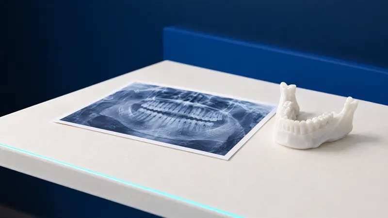
- A panoramic X-ray for an overview of all teeth and jawbones.
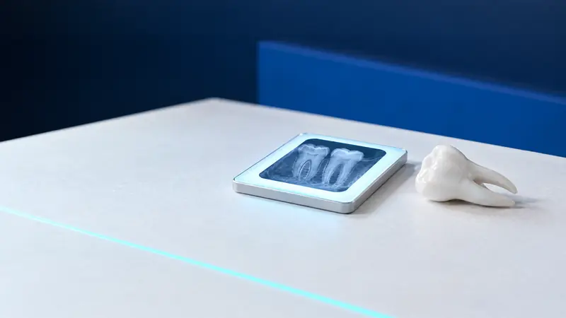
- Intraoral X-rays of teeth raising doubts on the panoramic image or examination.
- Small-field CBCT when findings are unclear and the result could change treatment.
Is a panoramic X-ray enough to exclude infection foci?
A panoramic X-ray is not always sufficient. It shows the whole dental arch in one image, but individual root lesions are better assessed on intraoral X-rays. The joint AAE and American Academy of Oral and Maxillofacial Radiology (AAOMR) 2015 statement identifies intraoral X-rays as the first investigation in endodontics; small-field CBCT is used selectively rather than routinely.
A 2016 meta-analysis by Leonardi Dutra et al., based on artificially created lesions, found greater diagnostic accuracy for CBCT than intraoral X-rays and no data confirming panoramic accuracy for root lesion detection. Panoramic imaging therefore provides an overview; doubtful teeth receive additional intraoral X-rays.
Radiation doses are small: European Commission Radiation Protection 172 guidance gives 2.7 to 24.3 µSv for a panoramic X-ray and below 1.5 µSv for a single intraoral image. Small- and medium-field CBCT gives 11 to 674 µSv and is therefore not a screening test. The article comparing CBCT and panoramic X-rays explains both methods.
What infection foci can a dentist find and how are they treated?
Management depends on the type of focus, tooth condition and planned procedure date. The table covers typical situations; the dentist and referring doctor decide the treatment needed before a particular operation.
| Possible focus | Detection | Usual management |
|---|---|---|
| Dead tooth with a root lesion | Vitality tests, intraoral X-ray, sometimes CBCT | Root canal treatment or extraction |
| Root-treated tooth with a root lesion | Comparison of current and earlier X-rays, CBCT if unclear | Healing review; if disease persists, retreatment, root-end surgery or extraction |
| Deep decay with pulpitis | Examination, pulp tests, bitewing X-ray | Pulp treatment or root canal treatment |
| Periodontitis with deep pockets | Pocket probing, X-rays | Periodontal treatment, extraction of teeth that cannot be retained |
| Wisdom tooth with recurrent gum inflammation | Examination, panoramic X-ray, CBCT if near a nerve | Extraction after the second or subsequent episode |
| Infected root left in bone | Panoramic and intraoral X-rays | Root removal after imaging assessment |
Dental clearance certificates in Białystok: how to obtain one and what does the NFZ cover?
A dentist issues a certificate stating there are no infection foci after examination and X-rays. Its wording depends on the referring doctor’s request. Ask beforehand whether a description of the teeth and periodontal health is enough or confirmation of completed treatment is also needed.
Treatment takes time, so arrange the dental appointment in advance. Before planned artificial heart valve implantation, ESC recommends eliminating infection sources at least 2 weeks beforehand; root canal treatment or several extractions may need multiple visits. An urgent procedure is the exception: do not postpone it yourself because of dental treatment.
The NFZ, Poland’s National Health Fund, entitles adults to one examination with oral hygiene instruction and three check-ups per calendar year, plus up to 2 intraoral X-rays annually. Panoramic X-rays are covered only for specified indications, with a referral, no more than once every 5 years. For adults without special entitlements, root canal cover is limited to tooth positions 1 to 3, while pregnant women and women in the postpartum period are entitled to root canal treatment of all teeth (legal status as of 24 October 2026).
The Podlaskie NFZ branch lists 206 general dental service locations and 16 oral surgery and periodontology locations in the region under contracts from 1 July 2025. The NFZ “Gdzie się leczyć” search and helpline 800 190 590 can identify a surgery in or around Białystok (as of 24 October 2026).
How it works at DentalBlue
At DentalBlue in Białystok, assessment for infection foci can begin with a “Wizyty bez bólu” (visit without pain), including a panoramic X-ray, consultation, examination and written treatment plan, priced at 300 zł. The clinic’s own radiology unit takes intraoral X-rays and CBCT scans. Root canal treatment and retreatment are performed under a microscope; the clinic also provides oral surgery and periodontology, so detected lesions can be treated on site. The clinician determines the final scope and cost after examination; prices may change.
Frequently asked questions
Is a painless dead tooth an infection focus?
It can be. The AAE explains that pulp death alone does not produce a root lesion until the canal becomes infected, but identifies a dead tooth as needing root canal treatment. ESE 2023 guidance recommends treatment once diagnosis is confirmed, even without pain.
Can a root-treated tooth be an infection focus?
Yes, if an inflammatory lesion persists around its root. A 2021 review by Tibúrcio-Machado et al. reports such a lesion in 39% of previously root-treated teeth. The dentist compares current and earlier X-rays and assesses symptoms: a healing lesion may need only monitoring, while persistent infection may need retreatment, root-end surgery or extraction.
Is CBCT needed for a dental clearance certificate?
Usually not. AAE and AAOMR recommend starting with two-dimensional X-rays and using small-field CBCT when findings are unclear and could change treatment. The dentist decides after examination and X-rays.
How much does assessment for infection foci cost?
Cost depends on the number of images needed and whether detected lesions require treatment. The clinic lists “Pantomogram” (panoramic X-ray) at 120 zł, “Zdjęcie wewnątrzustne” (intraoral X-ray) at 60 zł and “Tomografia komputerowa- szczęka lub żuchwa” (CT scan of the upper or lower jaw) at 300 zł. The clinician determines the final scope and cost after examination; prices may change.
Can oral infection foci cause heart disease?
Research shows an association but does not prove that a particular tooth will cause disease. The EFP and World Heart Federation 2020 report gives strong epidemiological evidence that periodontitis increases cardiovascular risk; ESC 2023 guidance recommends eliminating dental infection sources before artificial heart valve implantation.
Sources
- Int Endod J: Tibúrcio-Machado CS et al., global prevalence of apical periodontitis, meta-analysis (2021)
- J Calif Dent Assoc: Pallasch TJ, Wahl MJ, focal infection theory, appraisal and reappraisal (2000)
- J Clin Periodontol: Sanz M et al., periodontitis and cardiovascular diseases, EFP and WHF report (2020)
- European Heart Journal: ESC guidelines on infective endocarditis (2023)
- AAE: Colleagues for Excellence, Endodontic Diagnosis (2013)
- J Endod: AAE and AAOMR position statement on CBCT in endodontics (2015)
- Podlaskie NFZ branch: Leczenie stomatologiczne, dental service locations in Podlaskie (2026)
- ELI: Rozporządzenie MZ w sprawie świadczeń gwarantowanych z zakresu leczenia stomatologicznego, t.j. Dz.U. 2025 poz. 615
An informational article. It does not replace a consultation, and treatment decisions are made during a visit.